
Angewandte
Chemie
Metalloenzymes Hot Paper
Exploring Structure and Function of Redox
Intermediates in [NiFe]-Hydrogenases by an Advanced
Experimental Approach for Solvated, Lyophilized and
Crystallized Metalloenzymes
Christian Lorent,* Vladimir Pelmenschikov, Stefan Frielingsdorf,
Janna Schoknecht, Giorgio Caserta, Yoshitaka Yoda, Hongxin Wang,
Kenji Tamasaku, Oliver Lenz, Stephen P. Cramer,Marius Horch,*
Lars Lauterbach,* and Ingo Zebger*
In memory of Frank Koschine
A
ngewandte
Chemie
Research Articles
How to cite: Angew.Chem. Int. Ed. 2021,60,15854–15862
International Edition: doi.org/10.1002/anie.202100451
German Edition: doi.org/10.1002/ange.202100451
15854 T2021 TheAuthors.AngewandteChemieInternational Editionpublished by Wiley-VCHGmbH Angew.Chem. Int.Ed. 2021,60,15854–15862
15213773, 2021, 29, Downloaded from https://onlinelibrary.wiley.com/doi/10.1002/anie.202100451 by Cochrane Germany, Wiley Online Library on [29/12/2022]. See the Terms and Conditions (https://onlinelibrary.wiley.com/terms-and-conditions) on Wiley Online Library for rules of use; OA articles are governed by the applicable Creative Commons License
Abstract: To study metalloenzymes in detail, we developed
anew experimental setup allowing the controlled preparation
of catalytic intermediates for characterization by various
spectroscopic techniques.The in situ monitoring of redox
transitions by infrared spectroscopyinenzyme lyophilizate,
crystals,and solution during gas exchange in awide temper-
ature range can be accomplished as well. TwoO
2-tolerant
[NiFe]-hydrogenases were investigated as model systems.First,
we utilized our platform to prepare highly concentrated
hydrogenase lyophilizate in aparamagnetic state harboring
abridging hydride.This procedure proved beneficial for 57Fe
nuclear resonance vibrational spectroscopyand revealed, in
combination with density functional theory calculations,the
vibrational fingerprint of this catalytic intermediate.The same
in situ IR setup,combined with resonance Raman spectrosco-
py,provided detailed insights into the redoxchemistry of
enzyme crystals,underlining the general necessity to comple-
ment X-raycrystallographic data with spectroscopic analyses.
Introduction
Transition metals are often involved in chemical and
enzymatic catalysis.Innature,metal-containing enzymes
catalyze avariety of reactions,especially the conversion of
small gaseous molecules like CO2,N
2,orH
2.This type of
chemistry is relevant for establishing alternative strategies for
energy conversion and the production of carbon-neutral fuels.
Many of these metalloenzymes,including carbon monoxide
dehydrogenase,nitrogenase and hydrogenase,are attractive
targets for biotechnological application and can serve as
blueprints for bioinspired chemistry.[1–6] However,their
rational utilization requires athorough mechanistic under-
standing,typically requiring multiple spectroscopic tech-
niques.
Infrared (IR) spectroscopy in various implementations
has been successfully used to monitor catalytic processes and
intermediates at and beyond biological metal sites.Available
approaches comprise surface-sensitive techniques like sur-
face-enhanced infrared absorption (SEIRA) and/or attenu-
ated total reflection (ATR) spectroscopy,optionally com-
bined with electrochemistry,illumination, gas-atmosphere
and/or temperature control.[7–12] Recently,time-resolved IR
studies in the nanosecond range as well as ultrafast pump-
probe and two-dimensional IR techniques have also been
introduced into metalloenzyme research.[13,14] Electron para-
magnetic resonance (EPR) spectroscopy provides additional
structural and electronic insight into paramagnetic states,[15–18]
while resonance Raman (RR) spectroscopy has been success-
fully applied to monitor the characteristic metal@ligand
modes of specific redox states and cofactors.[19–24] Addition-
ally, 57Fe nuclear resonance vibrational spectroscopy (NRVS),
asynchrotron-based technique that can selectively probe
iron-specific normal modes,has been lately established for
characterizing iron-containing enzymes.[25–32]
Here,wehave designed anew experimental setup for
spectroscopic analyses of gas-converting metalloenzymes in
various sample forms.Gas composition and temperature can
be adjusted, thereby enabling the preparation of specific
redox states for subsequent characterization by complemen-
tary spectroscopic tools,such as RR, EPR, or NRVS.Addi-
tionally,redox transitions between catalytically relevant
intermediates and resting states can be monitored by in situ
IR spectroscopy.
To illustrate the versatility of our approach and its benefits
for exploring metalloenzyme catalysis,weinvestigated two
O2-tolerant model enzymes,the regulatory and the mem-
brane-bound [NiFe]-hydrogenase from Ralstonia eutropha
(ReRH and ReMBH, respectively). Briefly,[NiFe]-hydro-
genases catalyze the reversible cleavage of molecular hydro-
gen into protons and electrons (Figure 1). All of these
[*] C. Lorent, Dr.V.Pelmenschikov,Dr. S. Frielingsdorf,J.Schoknecht,
Dr.G.Caserta, Dr.O.Lenz, Dr.M.Horch, Dr.L.Lauterbach,
Dr.I.Zebger
Department of Chemistry,Technische Universit-tBerlin
Strasse des 17. Juni 135, 10623 Berlin (Germany)
E-mail:christian.lor[email protected]
Dr.Y.Yoda
Japan Synchrotron Radiation Research Institute
SPring-8
1-1-1 Kouto, Mikazuki-cho, Sayo-gun, Hyogo 679-5198 (Japan)
Dr.H.Wang, Prof. Dr.S.P.Cramer
SETI Institute
189 Bernardo Avenue, Mountain View,California 94043 (USA)
Dr.K.Tamasaku
RIKEN SPring-8 center
1-1-1 Kouto, Sayo-cho, Sayo-gun, Hyogo 679-5148 (Japan)
Dr.M.Horch
Department of Physics,Freie Universit-tBerlin
Arnimallee 14, 14195 Berlin (Germany)
E-mail:marius.horc[email protected]
Supportinginformation and the ORCID identification number(s) for
the author(s) of this article can be found under:
https://doi.org/10.1002/anie.202100451.
T2021 The Authors. Angewandte Chemie International Edition
published by Wiley-VCH GmbH. This is an open access article under
the terms of the Creative Commons Attribution License, which
permitsuse, distribution and reproduction in any medium, provided
the original work is properly cited.
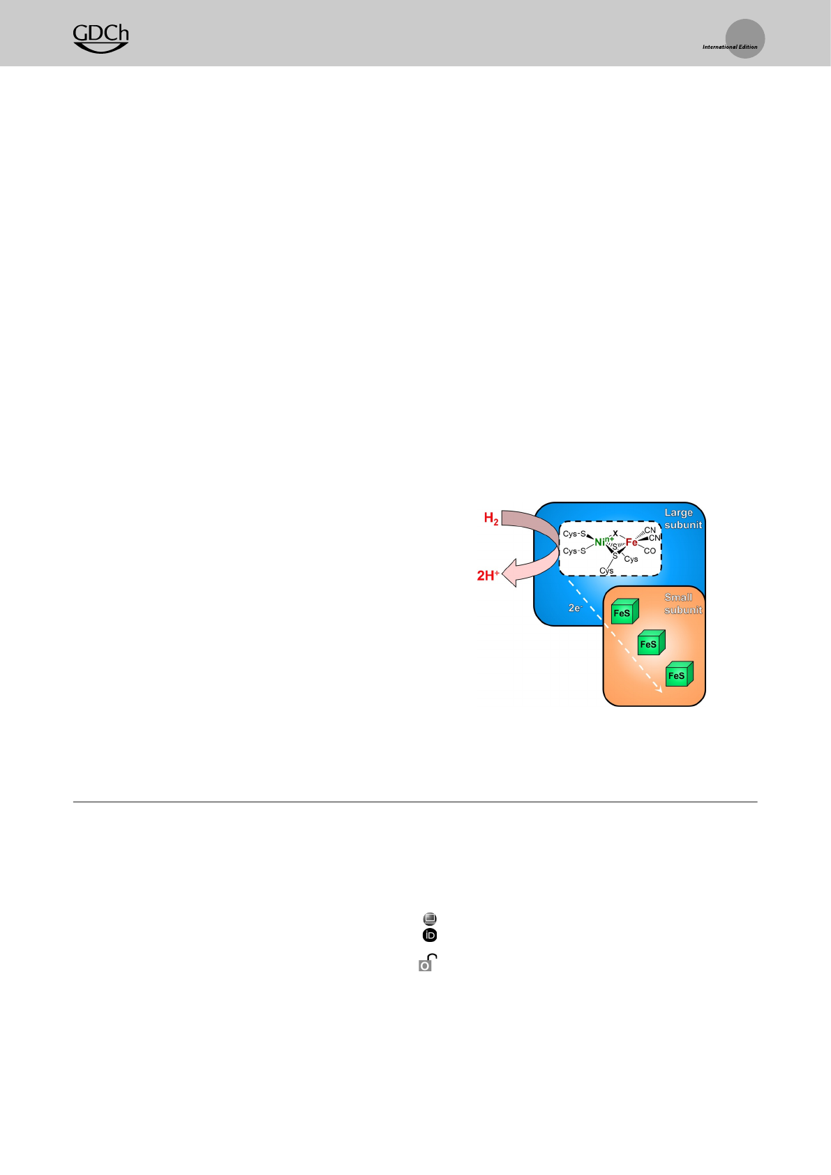
Figure 1. Schematic representationofthe consensusstructure and
cofactor composition of the model [NiFe]-hydrogenases investigated in
this study.The redox state of the active site is primarily determined by
the oxidation state of the nickel ion and the nature of the bridging
ligand X.
A
ngewandte
Chemie
Research Articles
15855Angew.Chem. Int.Ed. 2021,60,15854–15862 T2021 TheAuthors.AngewandteChemieInternational Editionpublished by Wiley-VCHGmbH www.angewandte.org
15213773, 2021, 29, Downloaded from https://onlinelibrary.wiley.com/doi/10.1002/anie.202100451 by Cochrane Germany, Wiley Online Library on [29/12/2022]. See the Terms and Conditions (https://onlinelibrary.wiley.com/terms-and-conditions) on Wiley Online Library for rules of use; OA articles are governed by the applicable Creative Commons License
enzymes feature aheterodimeric core structure,consisting of
alarge subunit harboring the [NiFe] active site and asmall
subunit comprising iron–sulfur clusters that form an electron
transfer relay.The proposed catalytic cycle and redox
intermediates of [NiFe]-hydrogenases discussed in this study
are displayed in Figure S1.
Results and Discussion
Theexperimental setup introduced here is designed for
the preparation of well-defined redox states of metalloen-
zymes and their concomitant multi-spectroscopic analysis
under various experimental conditions and in different
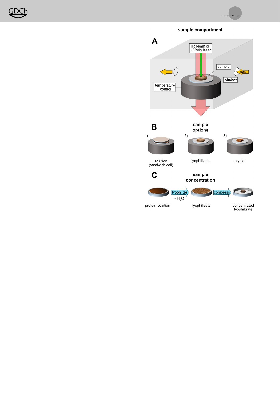
sample forms.The sample compartment consists of agas-
tight chamber connected to an external vacuum pump and
avariety of dry or humidified gases (Figures 2and S2). The
temperature can be set between 80 Kand 333 K. Our newly
developed approach also allows the in situ analysis of protein
solutions (Figures S3 and S11), lyophilizates (Figure 3C)and
single crystals (Figures 5and S10) by IR transmission
spectroscopy.Since the whole sample compartment is por-
table and gas-tight, it can also be disconnected from the
remaining setup and moved (Figure S2), for example,for RR
spectroscopic analysis of the same sample or further sample
treatment in an anaerobic chamber.
To illustrate the capabilities of our platform, we first
describe the preparation and spectroscopic characterization
of highly concentrated lyophilized samples of ReRH, suitable
for conducting NRVS experiments.Upon incubation with H2,
ReRH resides predominantly in the so-called Nia-C catalytic
intermediate.This species harbors abridging hydride between
the FeII and NiIII ions (Figure S1), which is of general interest
in biological and chemical catalysis.[33–35] So far, experimental
evidence for the presence of the hydride in this state has only
been provided by EPR spectroscopy.[35–37] Currently,the only
vibrational spectroscopic method suitable to monitor metal@
ligand bonding in this light-sensitive species (Figure S4) is
NRVS.[19,20] Observation of active-site metal@ligand vibra-
tions with this technique,however,typically requires protein
concentrations above 1mm,[27,28] which is the upper concen-
tration limit for purified ReRH in solution. To overcome this
limitation, we used our setup for agentle lyophilization to
prepare highly concentrated enzyme samples while preserv-
ing their catalytic activity.NRVSwas performed subsequently
in combination with density functional theory (DFT) calcu-
lations to reveal structural details of the Nia-C state.
In order to obtain highly concentrated samples in the Nia-
Credox state,0.5 mmprotein solution of ReRH was flash-
frozen at 77 Kand transferred to the sample compartment
(Figures 2A and S2). Subsequently,water was removed at
controlled temperature and pressure by applying amild
vacuum of 0.1 mbar while slowly warming up the sample to
243 K. After completion of the lyophilization, the sample
compartment was flushed with dry H2gas to reduce the
enzyme.Finally,compression of the protein lyophilizate,
using aspatula and apestle,yielded highly dense samples
(equivalent to 4–5 mm,see Supporting Information for de-
tails) suitable for demanding methods such as NRVS (Fig-
ure 2C). Forfurther details,see the Materials and Methods
section in the Supporting Information and Figure S2.
To verify structural integrity and functionality of the
lyophilized oxidized and H2-reduced enzyme,wemeasured
the H2-oxidation activity and applied EPR, IR and RR
spectroscopy (Figure 3). This multi-spectroscopic approach
was only applicable due to the design of the setup.The IR
signature of the amide Iand amide II vibrational modes is
related to the absorption of the (polyamide) backbone of the
protein and therefore reflects its secondary and tertiary
structure.[38] Thesimilarity between the spectra of the
lyophilized and solution-phase samples indicates that the
freeze-drying process did not affect the integrity of the
protein structure (Figure 3A). Additionally,lyophilized and
Figure 2. Setup for in situ IR analysis of proteins. A) The sample
compartment consists of agas-tight, temperature-controlled chamber
with IR-transparent windows. Agas supply allows the exchange of
various dried or humidified gases (H2,N
2,O
2)and mixturesthereof.
B) Protein samples can be studied in multiple forms:insolution in
asandwich cell made of two IR windows (Figures 5, S3B and S11B),
as lyophilized powder (Figures 3Cand S3A) or in the crystalline phase
(Figures 5and S11A) on top of asingle window.C)The concentration
of aprotein sample can be significantly increased, up to an equivalent
of 4–5 mm,via lyophilization and subsequent compression.
A
ngewandte
Chemie
Research Articles
15856 www.angewandte.org T2021 TheAuthors.AngewandteChemieInternational Edition publishedbyWiley-VCH GmbH Angew.Chem. Int.Ed. 2021,60,15854–15862
15213773, 2021, 29, Downloaded from https://onlinelibrary.wiley.com/doi/10.1002/anie.202100451 by Cochrane Germany, Wiley Online Library on [29/12/2022]. See the Terms and Conditions (https://onlinelibrary.wiley.com/terms-and-conditions) on Wiley Online Library for rules of use; OA articles are governed by the applicable Creative Commons License
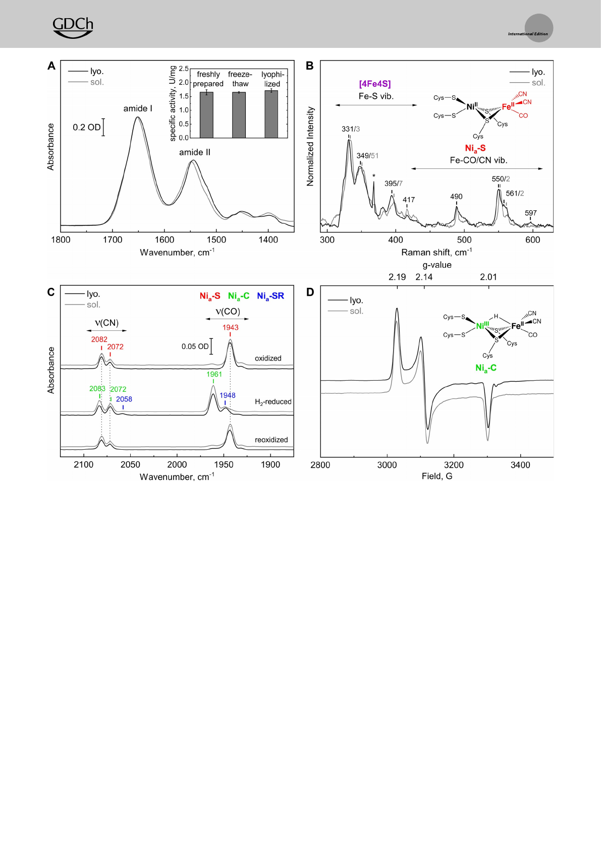
subsequently re-dissolved ReRH showed the same hydro-
genase activity as freshly isolated enzyme,indicating that the
metal cofactors and amino acid side chains responsible for
substrate conversion and proton/electron transfer were not
altered by lyophilization (Figure 3A inset). Theintegrity of
the iron–sulfur cluster relay was verified by almost identical
Fe@Ssignals in the corresponding RR spectra (Figure 3B).
Likewise,metal@ligand vibrations,characteristic for the most
oxidized, hydrogen-binding [NiFe] intermediate,termed Nia-
S(Figure S1), were observed, thereby confirming anative
active-site structure of oxidized ReRH lyophilizate (Fig-
ure 3B).[20]
Intriguingly,the lyophilized enzyme still reacts with
molecular hydrogen, as monitored by in situ IR spectroscopy
(Figure 3C). Thebiologically unusual CO and CN@ligands at
the active site of [NiFe]-hydrogenases give rise to valuable IR
marker bands that allow the identification of individual redox
states of the [NiFe] center.[39–41] TheH
2-dependent formation
of the Nia-C state indicates aredox-active ReRH in the
compressed and H2-reduced lyophilizate.Complementary
EPR spectra display the typical rhombic signature of the Nia-
Cstate,[35] confirming anative active-site structure after
lyophilization, reduction and compression (Figure 3D).
Thelyophilization procedure described above yielded
highly concentrated, 57Fe-enriched ReRH samples,which
Figure 3. Spectroscopic and biochemical characterization of lyophilized (lyo.) ReRH compared to freshly prepared enzyme solution (sol.). A) IR
spectroscopic signature reflecting the amide Iand II vibrational modes [38] of the oxidized enzyme. The inset shows the specific H2-oxidation
activity of freshly prepared, freeze-thawed and re-dissolved lyophilized samples. B) RR spectra of the oxidized enzyme obtained with 458 nm
excitationand normalizedtothe most intense active site signal at 550–552 cm@1.The small shifts of 1–2 cm@1likely reflect differences between
protein purified from cells grown with either 57Fe (lyo.) or iron with natural isotope distribution (sol.). The sharp signal marked with an asterisk
derives from an optical artefact of the Raman spectrometer.C)IRspectra of ReRH, oxidized (top), H2-reduced (middle) and subsequently re-
oxidized with air (bottom).For details about the assigned active-siteredox states, see Figure S1 and Table S1. D) EPR spectra of the H2-reduced
enzyme in solution and as compressed lyophilizate (both measured at 80 Kand with 1mWmicrowave power). IR and EPR spectra of the protein
solution are normalized to the intensity of the lyophilized sample.
A
ngewandte
Chemie
Research Articles
15857Angew.Chem. Int.Ed. 2021,60,15854–15862 T2021 TheAuthors.AngewandteChemieInternational Editionpublished by Wiley-VCHGmbH www.angewandte.org
15213773, 2021, 29, Downloaded from https://onlinelibrary.wiley.com/doi/10.1002/anie.202100451 by Cochrane Germany, Wiley Online Library on [29/12/2022]. See the Terms and Conditions (https://onlinelibrary.wiley.com/terms-and-conditions) on Wiley Online Library for rules of use; OA articles are governed by the applicable Creative Commons License
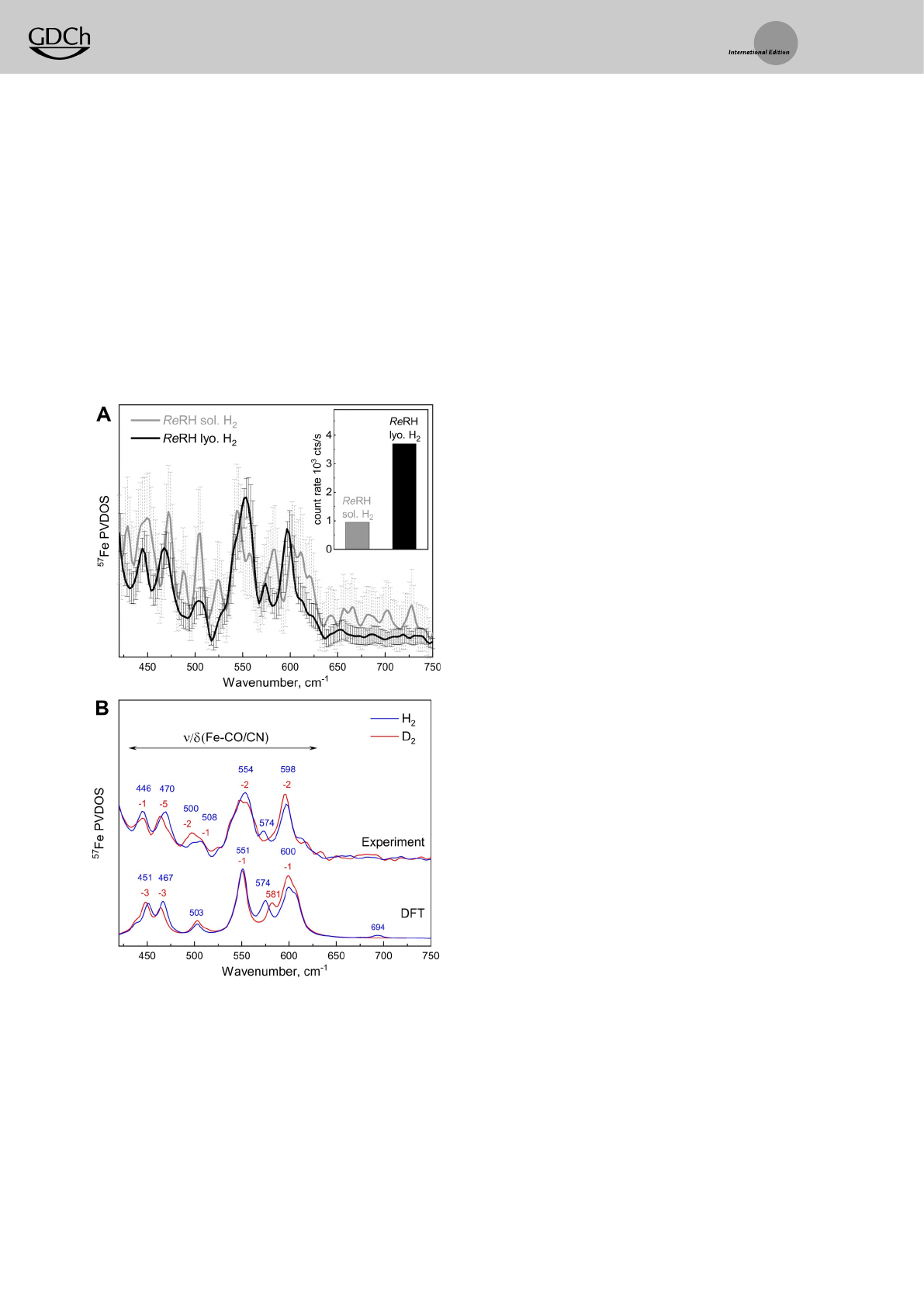
allowed NRVS-based detection of active-site vibrations of the
Nia-C state.The NRVS data shown in Figure 4comprise the
spectral regions characteristic for Fe@CO/CN modes (400–
650 cm@1)and Ni@H@Fe wagging vibrations (650–
800 cm@1).[25–28] Thecount rate of the elastic peak accumulated
by the detector increased by afactor of four for the
lyophilized sample in comparison to the solution-phase
sample (Figure 4A,inset), providing asignificant improve-
ment of the signal-to-noise ratio.Consequently,the error bars
of the two sample spectra vary,onaverage,byafactor of four
(Figure 4A,see Supporting Information for details).
In order to specifically probe the metal-bound hydride of
the Nia-C state,lyophilized ReRH samples were treated with
both H2and D2(Figure 4B). Theresulting experimental
spectra were compared to simulated data obtained by DFT
calculations (Figure 4B), based on an ReRH active-site
homology model[42,43] (Figures S5 and S6;for details,see the
DFT Methods section of the Supporting Information).
Analysis of the Nia-C spectrum obtained after incubation of
ReRH with H2(Figure 4B,top blue trace) revealed two
intense bands at 554 and 598 cm@1and aweaker one at
574 cm@1.Additional spectral features appear at 446, 470, 500
and 508 cm@1.Previously,[NiFe]-hydrogenase vibrational
signals at these energies were attributed to normal modes
dominated by Fe@CO and Fe@CN stretching and bending
motions.[19–21,25–28] Overall, the agreement between the exper-
imental and calculated spectra is good (Figure S7). The
isolated Ni@H@Fe wagging motions are predicted to produce
only very weak 57Fe-PVDOS intensities in the 670–740 cm@1
region. Themost noticeable spectral feature of this type is
expected at 694 cm@1and formed by two modes at 692 and
696 cm@1,each characterized by approximately 30% mH-
PVDOS but only subtle 1%57Fe-PVDOS.Adiscrete hydride
band could not be resolved by NRVS due to the low signal-to-
noise ratio in this spectral region, far away from the elastic
Mçssbauer peak. However,adistinct H/D-sensitive band
observed at 574 cm@1is predicted to arise from two modes
calculated at 574 and 576 cm@1(7% 57Fe-PVDOS each), both
of which are combinations of mHwagging and C@NH2
bending of the nearby Arg411 guanidium group (Figures S7A
and S8). TheNRVSdata recorded from ReRH incubated with
D2(Figure 4B,top red trace) reveal small shifts of 1–5 cm@1to
lower frequencies for several bands.This can be explained by
deuteride motions contributing to the Fe@CO(CN)-dominat-
ed modes,[20,44,45] with a mD-PVDOS maximum (23%) calcu-
lated at 501 cm@1for Nia-C(mD) (Figures S7B and S8, normal
mode animations provided in Supporting Information). Most
prominently,the 574 cm@1band detected for the H2-reduced
sample is absent in spectra from both the experimental D2-
treated sample and the calculated Nia-C(mD) model. More-
over, anewly emerging signal (calculated Nia-C(mD) mode at
581 cm@1)can be observed as aweak shoulder at ca. 586 cm@1,
close to the nearby high-intensity feature centered at
596 cm@1,indicating the presence of the mDdeuteride.In
summary,application of the novel setup proved to be
beneficial for the enrichment of ReRH, thereby enabling
NRVS characterization of the reduced enzyme.Rationalized
by DFT calculations,these data allowed insights into active-
site metal@ligand vibrations including normal modes that
reflect hydride coordination in the Nia-C redox state.
Using ReMBH as amodel metalloenzyme,wenext
demonstrate the applicability of our setup for the character-
ization of protein crystals by multiple spectroscopic tech-
niques (Figures 2B and S10). To obtain acomprehensive
understanding of redox processes in [NiFe]-hydrogenase
crystals,wefirst compared in situ IR spectra of aerobically
grown protein crystals and solution (Figure 5A,B). Unlike
Ash et al.,who employed electrochemical control in combi-
nation with redox mediators to induce redox transitions
within crystals of Hydrogenase 1from Escherichia coli,[46] our
design allows reduction of the hydrogenase with its native
substrate H2and (re)oxidation with the inhibitor O2.Thus,
our setup allows monitoring physiologically relevant redox
processes at the [NiFe] active site,asexemplified for the
Figure 4. Nuclear resonancevibrational spectra of ReRH in the Nia-C
state. A) NRVS data of lyophilized,H
2-reduced and compressed ReRH
(lyo.,black trace) compared to the corresponding protein solution at
0.5 mm(sol.,gray trace). Both spectra were recorded for 12 hours. The
inset displaysthe corresponding count rates at the elastic peak.
B) Experimental NRVS data of lyophilized ReRH (top) incubated with
H2(blue traces, 26 hours accumulation)orD
2(red traces, 16 hours
accumulation) in comparison to the corresponding DFT-calculated
57Fe-PVDOS spectra, which were obtained using the model shown in
Figure S6. NRVS data includingerror bars are displayed in Figure S9.
A
ngewandte
Chemie
Research Articles
15858 www.angewandte.org T2021 TheAuthors.AngewandteChemieInternational Edition publishedbyWiley-VCH GmbH Angew.Chem. Int.Ed. 2021,60,15854–15862
15213773, 2021, 29, Downloaded from https://onlinelibrary.wiley.com/doi/10.1002/anie.202100451 by Cochrane Germany, Wiley Online Library on [29/12/2022]. See the Terms and Conditions (https://onlinelibrary.wiley.com/terms-and-conditions) on Wiley Online Library for rules of use; OA articles are governed by the applicable Creative Commons License
Loading more pages...