Deterministic Generation and Guided Motion of Magnetic
Skyrmions by Focused He+‑Ion Irradiation
Lisa-Marie Kern, Bastian Pfau,*Victor Deinhart, Michael Schneider, Christopher Klose,
Kathinka Gerlinger, Steffen Wittrock, Dieter Engel, Ingo Will, Christian M. Gunther, Rein Liefferink,
Johan H. Mentink, Sebastian Wintz, Markus Weigand, Meng-Jie Huang, Riccardo Battistelli,
Daniel Metternich, Felix Buttner, Katja Höflich, and Stefan Eisebitt
Cite This: Nano Lett. 2022, 22, 4028−4035
Read Online
ACCESS Metrics & More Article Recommendations *
sıSupporting Information
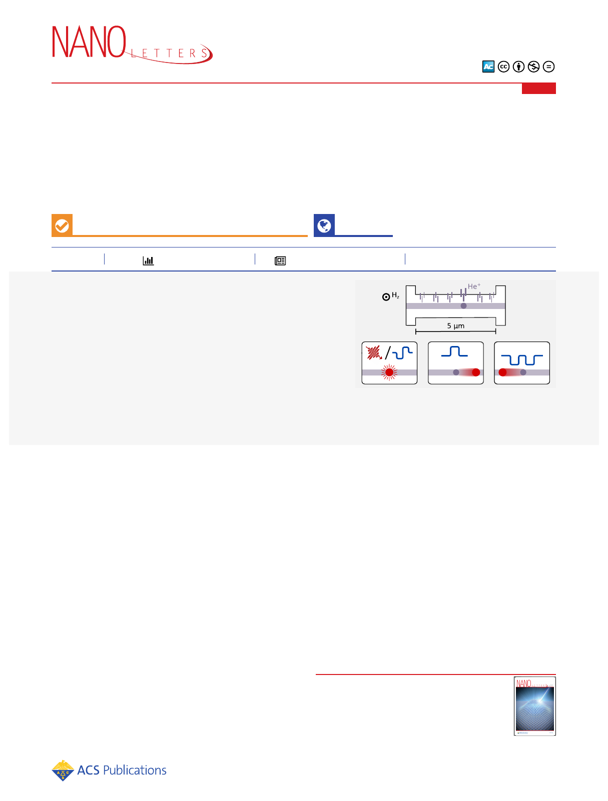
ABSTRACT: Magnetic skyrmions are quasiparticles with nontrivial top-
ology, envisioned to play a key role in next-generation data technology while
simultaneously attracting fundamental research interest due to their emerging
topological charge. In chiral magnetic multilayers, current-generated spin−
orbit torques or ultrafast laser excitation can be used to nucleate isolated
skyrmions on a picosecond time scale. Both methods, however, produce
randomly arranged skyrmions, which inherently limits the precision on the
location at which the skyrmions are nucleated. Here, we show that
nanopatterning of the anisotropy landscape with a He+-ion beam creates
well-defined skyrmion nucleation sites, thereby transforming the skyrmion
localization into a deterministic process. This approach allows control of
individual skyrmion nucleation as well as guided skyrmion motion with nanometer-scale precision, which is pivotal for both future
fundamental studies of skyrmion dynamics and applications.
KEYWORDS: magnetic skyrmions, ion irradiation, current-induced and laser-induced dynamics, magnetic racetrack, soft X-ray imaging
Magnetic skyrmions are topological quasiparticles that can
be as small as a few nanometers.
1−3
During the past
decade, research on magnetic skyrmions attracted interest from
scientific
4,5
and industrial
6
research communities due to the
skyrmion’s fascinating properties emerging from its topological
charge. They can exist in thin film materials with perpendicular
magnetic anisotropy (PMA) and are stabilized by stray fields
and the Dzyaloshinskii−Moriya interaction (DMI).
7−13
In
particular, skyrmions in chiral magnetic multilayer systems can
occur as isolated particle-like textures at room temperature and
in a low external magnetic field regime down to zero field.
13−15
Great advances have been reported in generating, annihilating,
and shifting skyrmions via spin−orbit torques (SOT) induced
by electric currents inside a suitable magnetic race-
track.
3,12,14,16−20
Moreover, recent studies have revealed that
faster and potentially more energy-efficient skyrmion gen-
eration is feasible by replacing current pulses with femtosecond
laser pulses, allowing optical nucleation even with a single laser
pulse.
21−23
While the underlying mechanisms are different, both
current-induced and optical nucleation suffer from a random-
ness in the spatial distribution of the skyrmions nucleated.
SOT magnetic switching and nucleation of a skyrmion are
mediated either by an applied symmetry-breaking in-plane
magnetic field
24,25
or by a locally modified anisotropy at a
magnetic defect site in combination with DMI.
16,18
Natural
magnetic defects caused by structural and chemical inhomo-
geneity can act as such nucleation sites, but they also influence
the SOT-driven skyrmion motion.
14,26
Since the density and
distribution of these defects depend on the growth process of
the material, the localization of the skyrmions remains poorly
controllable. In the case of the optical nucleation, skyrmions
emerge from spin fluctuations during a high-temperature phase
after the laser excitation. They finally appear randomly
distributed inside the illuminated area of the magnetic
film.
23,27
However, a controllable, reproducible, and reliable
localization of the skyrmion nucleation is often requiredboth
for future applications in data technology
28,29
and for
fundamental research, e.g., to realize repetitive pump−probe
experiments on skyrmion dynamics.
18
Previous attempts to control the localization of skyrmions
include structural patterning of the magnetic film with
notches
16,30,31
and discs
11
as well as the introduction of
defects
32,33
and nanopockets
34
to define positions for skyrmion
Received: February 17, 2022
Revised: May 2, 2022
Published: May 16, 2022
Letterpubs.acs.org/NanoLett
© 2022 The Authors. Published by
American Chemical Society 4028
https://doi.org/10.1021/acs.nanolett.2c00670
Nano Lett. 2022, 22, 4028−4035
Downloaded via TU BERLIN on August 9, 2023 at 09:53:49 (UTC).
See https://pubs.acs.org/sharingguidelines for options on how to legitimately share published articles.
nucleation. However, all of these methods include significant
structural or even geometrical modifications of the magnetic
racetrack and render an unhampered motion of the skyrmion
after creation difficult. Recent work also employed Ga+-ion
bombardment to create artificial defects as pinning sites for
skyrmions.
35,36
Due to the strong structural impact of these
heavy ions, it again remains an open question if a controlled
detachment of the skyrmion from the pinning site created in
this fashion is possible. In contrast, nanopatterning with a
focused He+-ion beam is suitable to prepare magnetic channels
for magnetic domain and skyrmion motion.
37
Light-ion
irradiation causes only minor structural reorganization on the
atomic scale, giving rise to an increased interfacial roughness
and intermixing of layerswithout affecting the topogra-
phy.
38−44
As a consequence, magnetic properties such as
coercivity, PMA, and DMI can be controlled at the
nanoscale.
40,41,43,45,46
In this work, we use a focused He+-ion beam to control the
localization of current-induced and laser-induced magnetic
skyrmions in ion-irradiated areas with different shapes and
sizes. In conjunction with the applied magnetic field, we can
turn the nucleation into a fully deterministic process
guaranteeing skyrmions to be generated in irradiated sites
while simultaneously suppressing their creation in non-
irradiated regions. Furthermore, we demonstrate controlled
SOT-induced detachment from such a nucleation site and the
subsequent guided motion of a single skyrmion over several
micrometers along a linear path, precisely defined by ion
irradiation, thereby effectively diminishing the transverse
skyrmion drift due to the skyrmion Hall effect. Our results
show the large potential of magnetic anisotropy patterning by
He+irradiation in multilayer structures for applied and
fundamental research on isolated skyrmions.
We prepared ferromagnetic multilayers with a nominal
composition of Ta(3 nm)/Pt(4 nm)/[Pt(2.5 nm/
Co60Fe25B15(0.95 nm)/MgO(1.4 nm)]15/Pt(2 nm) (see Sup-
porting Information for more details on sample fabrication).
The material system is well-known from previous experiments
on skyrmion nucleation and supports both SOT- and laser-
assisted nucleation.
16,23,27
He+-ion nanopatterning was carried
out with a ZEISS Orion Nanofab (acceleration voltage: 30 kV).
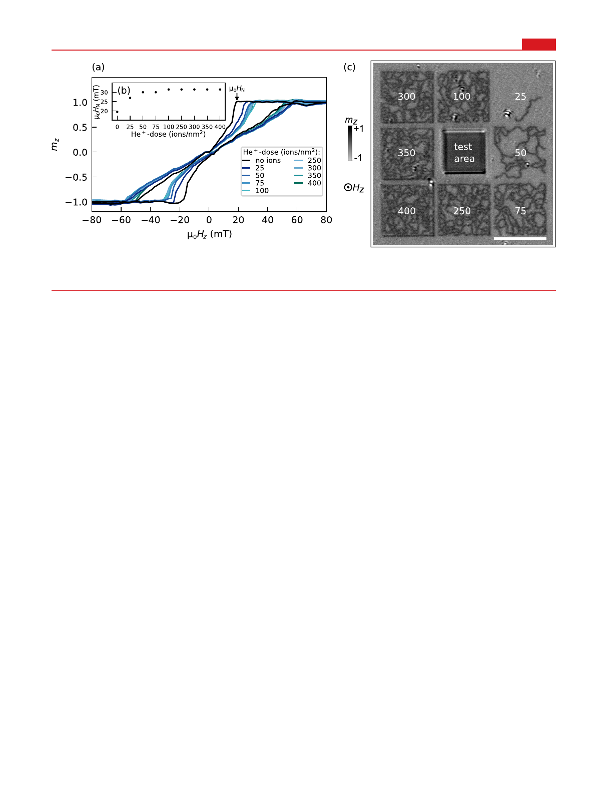
We locally irradiated the magnetic film with He+-ion doses
between 25 and 400 ions/nm2in predefined patterns with a
nominal resolution below 10 nm. However, the ion impact
diameter broadens to approximately 20 nm (full width at half-
maximum, fwhm) due to the ion collision cascade in our more
than 80 nm thick multilayer stack (see Supporting Information
for details). In Figure 1a, we present the magnetic hysteresis,
measured with Kerr microscopy on the pristine film and the
irradiated regions as shown in Figure 1c. In the irradiated
regions, at doses below 50 ions/nm2, the field HNallowing for
spontaneous nucleation increases with increasing dose. Above
50 ions/nm2, this dependence diminishes and no further
increase of HNis observed for ion doses above 100 ions/nm2
(see also Figure 1b). This behavior is in line with stopping and
range of ions in matter (SRIM) simulations for the parameters
in our experiment: The additional intermixing effect caused by
the He+-ions decreases with increasing ion dose and saturates
for ion doses above 100 ions/nm2(see Supporting
Information). The characteristic out-of-plane hysteresis shape
and the presence of labyrinth-like domains confirm that the
magnetic multilayer retains its perpendicular anisotropy and
supports magnetic domains in the nanometer range for all He+-
ion doses used here.
First, we present skyrmion nucleation in 2D-, 1D-, and 0D-
like ion-irradiated areas with homogeneous ion doses within
each pattern. We then turn our attention to the movement of
skyrmions in more complex irradiation profiles. Both the SOT
and laser-induced skyrmion nucleation were investigated in
samples patterned into a racetrack geometry
4,17
(Figure 2),
irradiated with He+-ions in different shapes and doses. We
imaged the magnetic textures nucleated via scanning trans-
mission X-ray microscopy (STXM) and Fourier-transform
holography (FTH) in transmission, utilizing X-ray magnetic
circular dichroism (XMCD) to obtain sensitivity to the
magnetization perpendicular to the sample plane (mz) (see
Supporting Information for more experimental details).
For the nucleation experiments, we first fully saturated our
magnetic film in an external magnetic field. In line with the
modification of the hysteresis behavior for different irradiation
doses, we reduced the magnetic field to a value for which
spontaneous nucleation of stripe domains remains suppressed
Figure 1. Magnetic hysteresis of He+-ion irradiated multilayer. (a) Normalized out-of-plane magnetization mzas a function of magnetic field Hzfor
variable He+-ion dose. (b) Nucleation field HNas a function of applied He+-ion dose. (c) Kerr image of irradiated areas in the magnetic film for
μ0Hz= 26 mT after saturation. Ion doses in ions/nm2. Scalebar corresponds to 10 μm.
Nano Letters pubs.acs.org/NanoLett Letter
https://doi.org/10.1021/acs.nanolett.2c00670
Nano Lett. 2022, 22, 4028−4035
4029
but skyrmion nucleation from an external stimulus is
possible.
16,27
At the outset, we confirmed that our non-
irradiated Pt/CoFeB/MgO multilayer supports skyrmion
nucleation. As a reference for the unmodified magnetic film
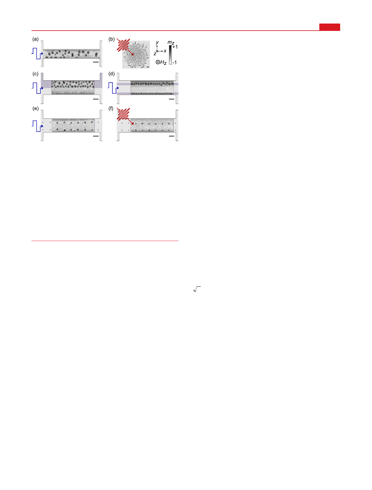
in Figure 2a and b, we present images of the skyrmion patterns
created by applying a single bipolar current pulse (jx=±1.1 ×
1012 A/m2) and a single infrared (IR) laser pulse (53 mJ/cm2),
respectively (see Supporting Information for pulse parame-
ters). For the nonirradiated magnetic film, we observed a
random distribution of skyrmions in both cases. The reference
sample in Figure 2b was not patterned into a racetrack, and
skyrmions form in the entire area illuminated by the laser
where the laser fluence overcomes the nucleation thresh-
old.
23,27
In samples with an ion irradiated 2D area covering half
the racetrack (c) and with a 1D line pattern (d), we applied a
single bipolar current pulse to create skyrmions ((c) jx=±1.3
×1012 A/m2, (d) jx=±7.7 ×10¹¹ A/m2). To suppress
nucleation in nonirradiated regions, we increased the applied
field compared to the nucleation in nonirradiated racetracks. In
this fashion, skyrmion nucleation only occurs in irradiated
regions. Within these extended 2D and 1D ion-irradiated areas,
the skyrmions preserve their spherical shape compared to the
pristine film and appear almost homogeneously distributed. In
Figure 2e and f, we show the results from current-induced and
optical excitations in racetracks with predefined 0D dot
patterns (100 nm diameter) by He+-ion irradiation ((e) jx=
±7.7 ×10¹¹ A/m2, (f) 45 mJ/cm2). For appropriate external
fields, we observe skyrmion nucleation exclusively and exactly
in the predefined dots.
In Figure 1 and Figure 2, we demonstrated that we can
employ the applied field to effectively suppress nucleation in
nonirradiated regions. Next, we discuss the influence of the
magnetic field on the skyrmion formation inside the predefined
regions. For He+patterned dots of 100 nm diameter, we
observed reliable nucleation at the dots by both laser pulses
and SOT pulses, as shown in Figure 3a and b, respectively.
Skyrmions nucleate at all dots, and exclusively there, over a
broad range of fields applied. Similar to skyrmions in
homogeneous media, the skyrmion diameter continuously
decreases with increasing magnetic field. The skyrmion size is,
thus, not fixed to the size of the He+irradiated area. In
contrast, for the skyrmion nucleation at He+patterned dots of
300 nm diameter (Figure 3c), we observe the generation of
noncompact magnetization textures. At low magnetic field, a
single bipolar current pulse creates a variety of almost closed or
fully closed ring-shaped domains (so-called skyrmionia
47
or
target skyrmions
48
) surrounding the irradiated dot. Increasing
the magnetic field mostly leads to the formation of a single
circular skyrmion per irradiated dot. At even higher applied
fields, several magnetic skyrmions (mostly three) stabilize
inside the He+-ion irradiated areas. We infer that the most
suitable choice for controlled nucleation of individual sky-
rmions is to rely on He+-ion irradiated dots of a size
comparable to or smaller than the skyrmion diameter
supported by the material at a particular field. Nevertheless,
larger He+dots provide an interesting route to create and
investigate topological textures beyond charge-unity sky-
rmions.
47
In Figure 3d, we present the skyrmion diameter on the array
of 100 nm sized He+-ion irradiated dots for different applied
ion doses in a range from 25 to 350 ions/nm2and as a function
of the external field. The current and optical excitation
amplitudes were chosen such that, for the lowest magnetic field
investigated, all predefined dots were filled with skyrmions
while nucleation in nonirradiated regions remains suppressed.
The excitation setting per sample was kept constant while
varying the magnetic field. The skyrmion diameter was
determined from the full width at half-maximum (fwhm) of
the central autocorrelation peak of the STXM images as fwhm/
2to account for the skyrmions’self-convolution. Each data
point thus corresponds to the average diameter of several
skyrmions recorded in one STXM image. In Figure 3d, we
observe a decrease in skyrmion diameter with increasing field
in the direction opposite to the net perpendicular magnet-
ization of the skyrmion from about 155 nm at 25 mT to 75 nm
at 52 mT. We attribute the large scatter to a spatial variation of
anisotropy and DMI and a variation of the location of the
irradiated regions within the narrowed racetrack affecting stray
fields.
49
Within this variance, there is no apparent systematic
dependence of skyrmion diameter on ion dose.
For many applications, the subsequent detachment of a
skyrmion from its predefined nucleation site will be important.
In the following, we will thus discuss the possibility for
detachment and guided motion after deterministic nucleation.
Afirst detachment experiment is presented in Figure 4,
performed via FTH-based imaging with a field of view (FOV)
of 1.2 μm diameter. We started by applying a single bipolar
current pulse (jx=±1.4 ×10¹² A/m2) to nucleate skyrmions
on a He+-ion irradiated dot array with 60 nm dot diameter. We
first imaged the nucleation from saturation five times to
demonstrate a reliable skyrmion formation in the irradiated
dot. While the skyrmion nucleated at the central dot is of
interest (Figure 4a), a second skyrmion is nucleated at a
neighboring dot visible in our FOV. In order to detach and
move the central skyrmion via SOT to the left, we applied a
Figure 2. Localized current-induced and optical skyrmion nucleation.
(a−b) Stochastic nucleation in the pristine material either by a
current pulse (a, current density jx=±1.1 ×10¹²A/m2,μ0Hz=26
mT) or by an optical laser pulse (b, peak fluence 53 mJ/cm2,μ0Hz=
40 mT). In (c−f), a scheme of the structure indicating the He+
irradiated regions (purple) has been overlaid with images of the out-
of-plane magnetization obtained by STXM. (c−d) Current-induced
nucleation in an irradiated area (c, 100 ions/nm2dose, jx=±1.3 ×
1012A/m2,μ0Hz= 27 mT) and on lines (d, 200 nm width, 350 ions/
nm2dose, jx=±7.7 ×10¹¹A/m2,μ0Hz= 40 mT). (e) Current-
induced nucleation in a dot array of dose 350 ions/nm2(jxas in (d),
μ0Hz= 47 mT). (f) Optical nucleation in a dot array of dose 350
ions/nm2(peak fluence 45 mJ/cm2,μ0Hz= 51.5 mT). (a−f)
Skyrmions are created from a single current or laser pulse. Scalebars
correspond to 500 nm.
Nano Letters pubs.acs.org/NanoLett Letter
https://doi.org/10.1021/acs.nanolett.2c00670
Nano Lett. 2022, 22, 4028−4035
4030
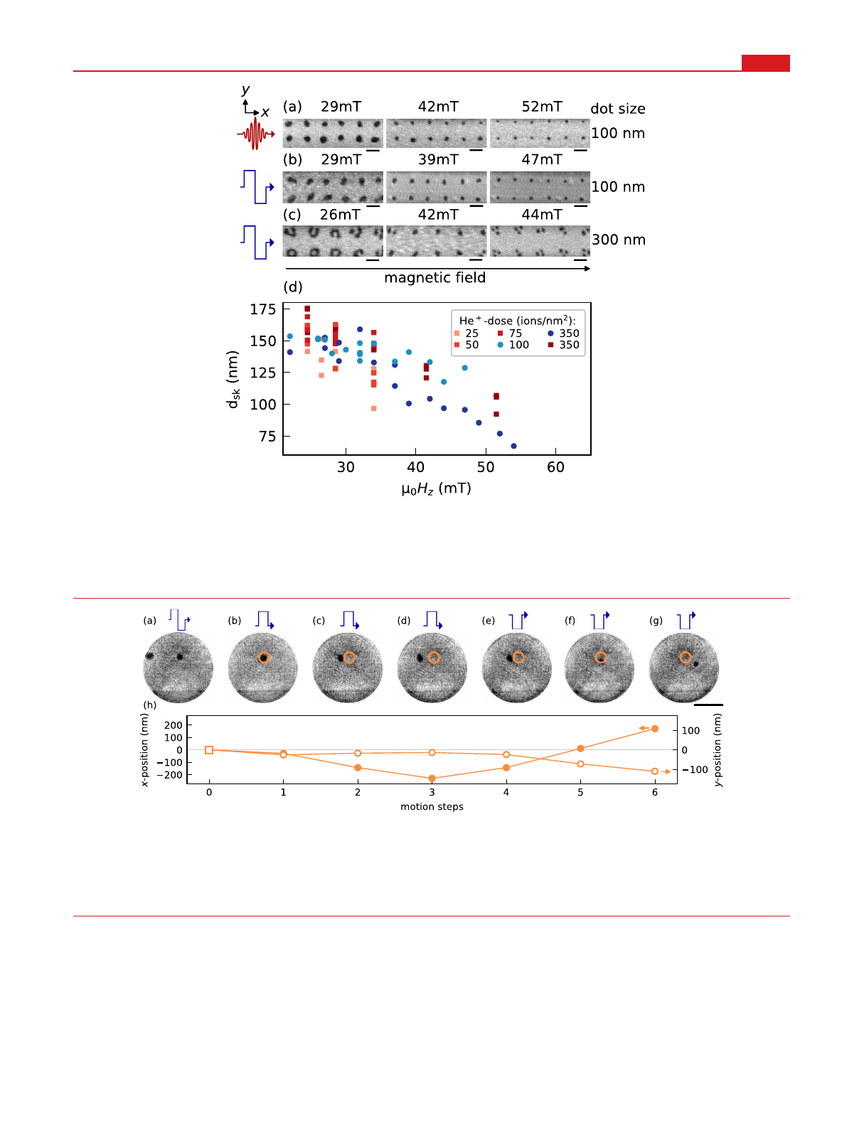
sequence of single unipolar current pulses of reduced current
density (jx= 4.3 ×10¹¹ A/m2) as compared to the nucleating
bipolar pulse and recorded an image after every pulse (Figure
4b−g). With the first unipolar pulse, the central skyrmion
mostly deforms which we interpret as being part of the
depinning from the nucleation site. Once detached, the
skyrmion can move freely back and forth. After three unipolar
current pulses (Figure 4b−d), we reversed the current
direction and the skyrmion moves in the opposite direction
(Figure 4e−g). Important in the context of device applications,
the magnetic field is kept constant during the entire sequence
including nucleation, detachment, and motion. Tracking the
position of the skyrmion in the x- and y-direction (Figure 4h),
we observe almost uniform motion steps along the current
direction with an average velocity of vx= 11(3) m/s. The
typical inclination of the motion path owing to the skyrmion
Hall effect
19,20
directly proves the topological nature of the
magnetic textures createdthey are skyrmions.
Figure 3. Magnetic field impact on skyrmion diameter and nucleation reliability. (a−c) STXM images after skyrmion nucleation in magnetic
racetracks with He+patterned dot arrays (350 ions/nm2dose). Nucleation method (symbols on the left), applied field (above each image), and
irradiated dot diameter (right) are indicated. Scalebars correspond to 500 nm. (d) Skyrmion diameter dsk as a function of the applied field in
magnetic magnetic racetracks with He+patterned arrays of dots with 100 nm diameter. Data points with red color shades and squared markers
correspond to laser-induced nucleation, and those with blue shades and round markers correspond to current-induced nucleation.
Figure 4. Controlled detachment from a He+-ion irradiated dot. (a−g) X-ray holography images of a skyrmion detaching from and moving over its
nucleation site due to single current pulses at constant applied field of μ0Hz= 36 mT. (a) Current-induced nucleation at predefined dots (50 ions/
nm2dose, jx=±1.4 ×10¹² A/m2), (b−g) detachment and free motion (jx= 4.3 ×10¹¹ A/m2). The orange circle marks the position of the
predefined nucleation site for the skyrmion of interest. A movie of the series is available as Supporting Information. The skyrmion at the edge of the
field of view is presumably moved out of the short racetrack (2 μm length) in the course of the motion to the left and then pinned in a region with
lower current density. Scalebar corresponds to 500 nm. (h) Distance traveled in x- and y-direction as indicated by the arrows.
Nano Letters pubs.acs.org/NanoLett Letter
https://doi.org/10.1021/acs.nanolett.2c00670
Nano Lett. 2022, 22, 4028−4035
4031
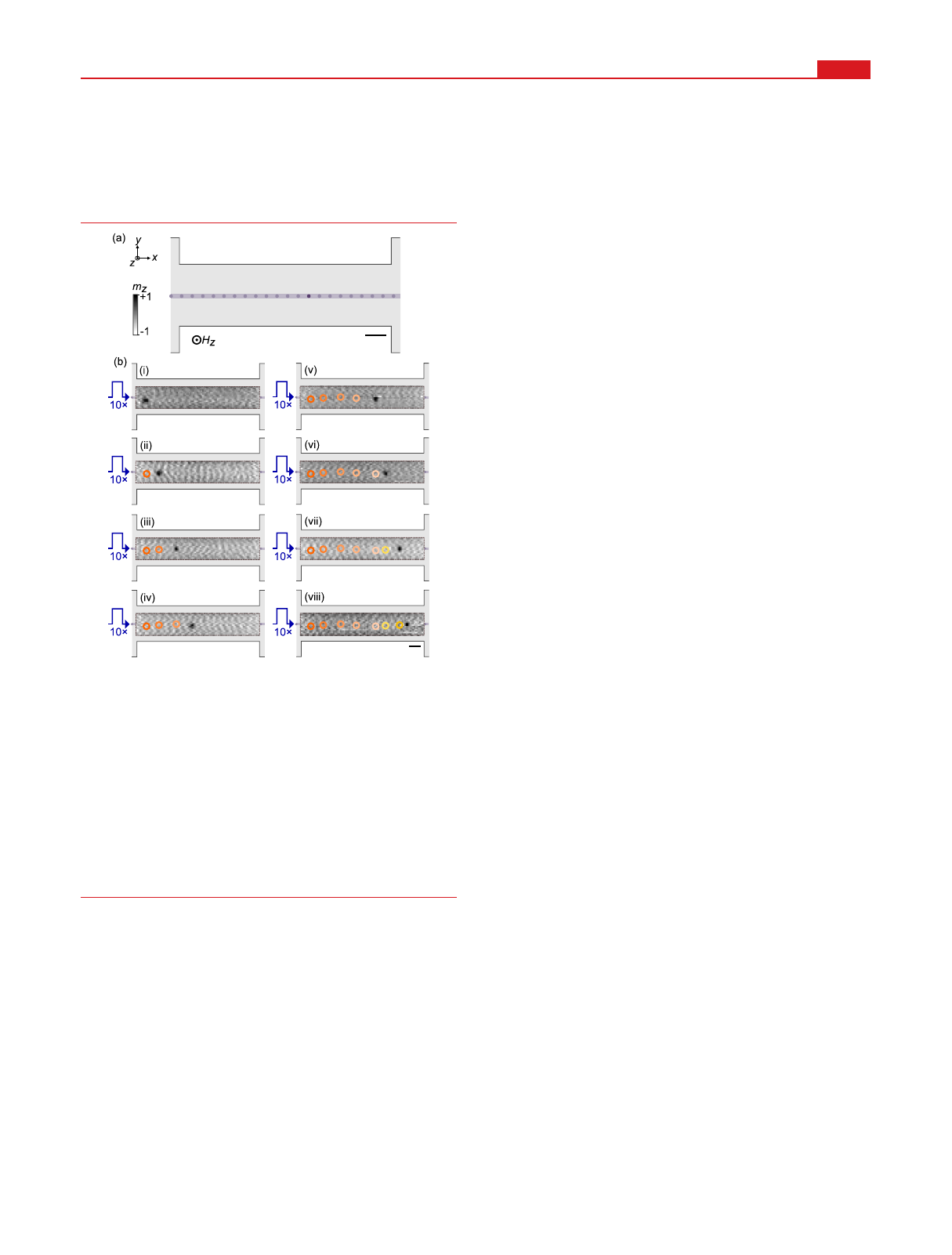
Next, we address the need to guide the SOT-driven
skyrmion motion within the racetrack in a fashion compatible
with the deterministic nucleation and subsequent detachment
demonstrated so far. To this end, we prepare a He+irradiation
pattern of dots (60 nm diameter, 50 ions/nm2), linked via 250
nm long connecting lines (25 ions/nm2), as illustrated in
Figure 5a. A single dot on the channel near the center of the
racetrack was produced with a higher dose of 100 ions/nm2to
act as a nucleation site. At this location, a single skyrmion
nucleates from a single bipolar current pulse (jx=±1.2 ×10¹²
A/m2) after saturation (not shown). Within the irradiated
channel, we moved the skyrmion with unipolar current pulses
(jx= 7.7 ×10¹¹ A/m2) at an average velocity of vx= 2.5(8) m/
s. We are able to propagate the skyrmion back and forth over
several micrometers along a straight line, covering the entire
length of the magnetic racetrack. For nucleation and SOT-
driven motion, a constant magnetic field of 39 mT was applied.
Figure 5b illustrates the guided motion with a selection of eight
(i−viii) consecutive STXM images of the motion series. The
full series including the nucleation at the central dot and all 24
motion steps is provided as a movie in the Supporting
Information. The skyrmion moves strictly along the guiding
channel; deviations in y-direction are smaller than the
skyrmion diameter of about 125 nm. Note that the skyrmion
also passes across its previous nucleation site without being
trapped when moving back and forth. Our approach also
directly supports the controlled synchronized motion of a train
of skyrmions as we present in Figure S2 of the Supporting
Information. The demonstration of this shift operation on a
whole bit pattern represents a crucial step toward proposed
memory applications of skyrmions.
4
Our findings prove that He+-ion irradiation represents an
excellent tool to reliably and precisely localize current-induced
and optical nucleation of isolated skyrmions in a magnetic
multilayer material. On the one hand, the magnetic
modification is effective enough to promote nucleation in
irradiated areas with full reliability while nucleation in
nonirradiated areas can be suppressed completely. On the
other hand, the modification is gentle enough that the
magnetic racetrack remains topographically intact and the
magnetic properties in the irradiated regions are preserved in
the sense that stable nanometer-scale skyrmions are formed,
with a size and shape that we cannot distinguish from
skyrmions in nonirradiated areas of the sample. The latter
finding strongly suggests that a local pinning potential possibly
created by ion irradiation can only play a minor role for the
localization of the nucleation.
Instead, recent work reported that ion irradiation can
significantly enhance the efficiency of SOT-driven magnet-
ization switching in heterostructures containing a single
ferromagnetic layer.
43,44,50
The reasons were found in a
reduced PMA of the irradiated structure
43,50
and an increased
spin-Hall angle from the Pt probably due to an increased defect
density in this layer.
44
In addition, intermixing at the Co−Pt
interfaces can lead to a higher spin transparency
51
and again to
an increased spin-Hall angle.
52
We expect that similar effects
are key also for the easier SOT nucleation of skyrmions in He+
irradiated areas of our material even though it is much thicker
compared to material systems used in refs 43,44, and 50. Our
SRIM simulations confirm a mostly homogeneous ion
penetration along the depth direction of our multilayer (see
Supporting Information). Consequently, the more efficient
SOT switching allows us to act against a significantly higher
applied field compared to the SOT in nonirradiated regions. In
the case of laser-induced skyrmion nucleation, we also
identified the reduced PMA in the He+irradiated areas as
main origin for the preferred skyrmion nucleation. We required
a reduced laser fluence to nucleate skyrmions in the ion-
irradiated dots. We modeled this dependence in atomistic
simulations of the high-temperature fluctuation phase which
leads to the formation of skyrmions
23
in model systems with
different values of the PMA. For the low-PMA material, the
simulations predict a lower excitation threshold for skyrmion
nucleation (see Supporting Information). The laser fluence,
thus, provides a handle to trigger the skyrmion nucleation
exclusively in the predefined regions.
In the second part of our work, we demonstrated the
localized nucleation of a skyrmion combined with its
detachment from the site and subsequent guided motion in a
narrow ion-irradiated channel. Such a controlled detachment
from the nucleation site and the unimpeded skyrmion motion
across it are only possible if the pinning potential formed by
the ion irradiation is sufficiently shallow and tunablean
important advantage of light ions over the heavier ions such as
Ga+that are routinely used for topographic modifications on
the same length scale. Our method to create skyrmion
Figure 5. Guided skyrmion motion. (a) Schematic irradiation layout
showing the nucleation dot (dark purple), the guiding channel (light
purple) with intermediate medium-dose dots. (b) STXM images of a
motion series at constant field of μ0Hz= 39 mT. After nucleation with
a single bipolar pulse at the nucleation dot (jx=±1.2 ×10¹² A/m2),
the skyrmion is imaged along the guiding chain in 24 steps. Eight
representative consecutive images are shown (i−viii). In order to
increase the distance moved between two images, ten consecutive
unipolar current pulses (jx= 7.7 ×10¹¹ A/m2) were applied before
recording the next image. The skyrmion moves back and forth along
the entire field of view with no systematic deviation from the
irradiated channel, i.e., with suppressed skyrmion Hall effect. The
orange-shaded circles mark previous positions. Scalebars correspond
to 500 nm.
Nano Letters pubs.acs.org/NanoLett Letter
https://doi.org/10.1021/acs.nanolett.2c00670
Nano Lett. 2022, 22, 4028−4035
4032
Loading more pages...