
Cite this: Lab Chip, 2013, 13, 3555
Skin and hair on-a-chip: in vitro skin models versus ex
vivo tissue maintenance with dynamic perfusion3
Received 19th February 2013,
Accepted 1st May 2013
DOI: 10.1039/c3lc50227a
www.rsc.org/loc
Beren Ataç,{
a
Ilka Wagner,{
a
Reyk Horland,
a
Roland Lauster,
a
Uwe Marx,
a
Alexander
G. Tonevitsky,
b
Reza P. Azar
c
and Gerd Lindner*
a
Substantial progress has been achieved over the last few decades in the development of skin equivalents
to model the skin as an organ. However, their static culture still limits the emulation of essential
physiological properties crucial for toxicity testing and compound screening. Here, we describe a
dynamically perfused chip-based bioreactor platform capable of applying variable mechanical shear stress
and extending culture periods. This leads to improvements of culture conditions for integrated in vitro skin
models, ex vivo skin organ cultures and biopsies of single hair follicular units.
Introduction
The majority of current commercially available skin equiva-
lents are based on static culture systems emulating human
epidermis only, or combining epidermis and dermis in so-
called full-thickness skin equivalents (for reviews, see
1,2
). None
of the existing systems comprise functional and (patho-)
physiologically important elements, such as an immune
system, vasculature or skin appendices.
3
Most of the skin-
related pathologies, such as wound healing, skin tumours,
psoriasis, contact allergies, androgenic alopecia, etc., essen-
tially require the elements mentioned above. Consequently,
reproducible in vitro systems should reflect these circum-
stances.
Further bioengineering is particularly necessary for the
implementation of adipose tissue, hair follicles and a
functional vascular network.
4–7
Notably, adipose-derived stro-
mal cells are not only important for lipid metabolism, but also
have a major impact on the regulation of fibroblast and
keratinocyte proliferation by cytokine secretion.
8
In addition,
the hair follicle serves as a considerable storage area, takes a
substantial role in skin metabolism, contains multiple stem
cell lineages with regenerative capacity, and constitutes the
major penetration route of topically applied substances to the
skin.
9–11
Native skin is supplied through a capillary network in the
dermis, formed by endothelial cells. Therefore, endothelial
cells have been integrated into the dermal portion of full-
thickness skin substitutes.
12,6,7
However, because of the lack
of shear stress in existing in vitro systems, no mature blood
vessels have been produced and no skin substitute with an
artificial blood stream at micro-scale has been described so
far.
Such an improved skin equivalent needs to be implemen-
ted in a culture system which permits a constant oxygen and
nutrient supply, measures and removes toxic metabolites, and
can be intravitally monitored for sustainability over weeks and
even months. This is necessary when it is used, for example, as
a potential replacement of animal studies for repeated dermal
toxicity assays, as required in OECD (Organisation for
Economic Co-operation and Development) guidelines 410
and 411. We have recently successfully developed a PDMS-
based microfluidic chip system with the dimensions of a
conventional glass microscope slide
13,14
to function as a long-
term dynamic bioreactor for various tissues. In this study, we
used the chip-based system to prolong the maintenance and
testing period of a commercially available skin equivalent (SE)
and improve its nutritional and cellular deposit with ex vivo
subcutaneous tissue (SCT). Furthermore, ex vivo skin and
single hair follicular units (follicular unit extracts – FUEs) were
cultured in the bioreactor platform to extend the static
maintenance periods concerning substance testing.
Materials and methods
Device design and assembly
The bioreactor platform we developed (Fig. 1a), which will
subsequently be referred as the ‘‘MOC’’ (Multi-Organ-Chip),
provides a dynamically perfused micro-channel system com-
a
Technische Universita
¨t Berlin, Institute of Biotechnology, Berlin, Germany.
E-mail: gerd.lindner@tu-berlin.de; Fax: +49 (0)30-314-27914;
Tel: +49 (0)30-314-27910
b
Institute of General Pathology and Pathophysiology Russian Academy of Medical
Science, Moscow, Russia
c
Zentrum fu
¨r moderne Haartransplantation / Centre for Modern Hair
Transplantation, Berlin, Germany
3
Conflict of interest disclosures: the authors declare no competing financial
interests.
{These authors contributed equally to this work.
Lab on a Chip
PAPER
This journal is ßThe Royal Society of Chemistry 2013 Lab Chip, 2013, 13, 3555–3561 |3555
Published on 01 May 2013. Downloaded by TU Berlin - Universitaetsbibl on 29/03/2016 10:22:02.
View Article Online
View Journal
| View Issue
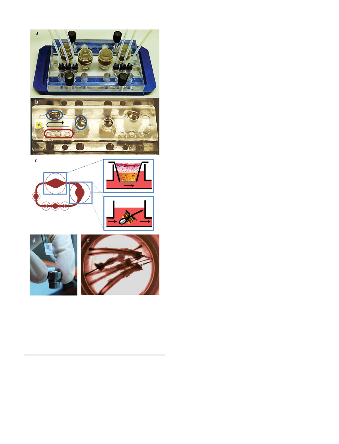
bining an on-chip micropump (Fig. 1b, red circle) with variable
tissue culture compartments (Fig. 1b, blue circle, Fig. 1c). The
MOC system provides a platform to assess the effect of
dynamic perfusion in comparison to common static culture
conditions of different skin model systems. It is designed to
operate two microfluidic circuits simultaneously. Fig. 1b is a
photo of the MOC showing the positioning of the key elements
at a glance. The layout of the MOC supports the flexible
integration of conventional miniaturised tissue culture for-
mats, such as Transwell1(Corning, Lowell, MA, USA) inserts,
special organotypic matrices, or native normal or diseased ex
vivo biopsies in the tissue culture compartments (Fig. 1c blue
rectangles). The micropump is capable of providing a pulsatile
flow of medium through 500 mm wide 6100 mm high
channels with a pumping volume range of 7–70 ml min
21
and a
frequency of 0.2–2.5 Hz. We used a frequency of 0.3 Hz in our
experiments, which leads to a flow-rate of approximately 30 ml
min
21
. SEs and skin biopsies (SBs) were each held in a single
96-well Transwell1insert with a surface area of 14.3 mm
2
,
placed in the insert holders (Fig. 1d) to provide an air–liquid
interphase (Fig. 1c), while FUEs were positioned directly into
the insert areas leading to submerged culture conditions
within the medium stream (Fig. 1c and e). For a detailed
description of the MOC assembly procedure, please refer to
the work of Wagner and colleagues in this issue.
15
Tissue sources and culture conditions
We used EpiDermFT
TM
(Mattek, Ashland, MA, USA) as a full-
thickness in vitro model for the in vitro SE experiments. A 5
mm punch biopsy device (Stusche, Teltow, Germany) was used
to adjust the SE’s size to the Transwell1(Corning, Lowell, MA,
USA) insert. Dulbecco’s Modified Eagle’s Medium (DMEM)-
based maintenance medium (EFT-400-MM, Mattek, Ashland,
MA, USA) was used, according to the manufacturer’s instruc-
tions. A total volume of 500 ml medium was in place, while 200
ml was changed every day during the 9 days of culture for both
MOC-based dynamic and static cultures in air–liquid inter-
phase. We used deep Transwell1holder wells for the static
cultures, with the same medium volume in all parallel
cultures.
Human juvenile prepuce was obtained in compliance with
the relevant laws, with informed consent and ethical approval
from the Ethics Committee of the Charite
´Universita
¨tsmedizin,
Berlin, Germany, after routine circumcisions from paediatric
surgery. The tissue was stored in PBS at +4 uC and processed
within 4 h after surgery. Skins were sterilized in 80% ethanol
for 30 s and cut open. SCT was removed from the skin and
placed underneath the SE, prepared as described above. These
combined tissues were positioned in the Transwell1insert
and transferred to the respective MOC compartment utilising
the same experimental conditions as mentioned above for a
duration of 7 days (Fig. 1d).
Similarly, we used juvenile prepuce for the ex vivo skin
experiments and processed them as described above. Three
donors were used for each MOC-based static and dynamic
experiment in parallel. The skin was separated from the
subcutaneous tissue, punched to 5 mm biopsies, fixed in the
Transwell1insert, and transferred to the MOC tissue segment
in air–liquid interphase. SBs were supplemented for 14 days in
high glucose (4.5 mg ml
21
) DMEM with 10% foetal calf serum
(FCS), 100 units ml
21
penicillin, 100 mgml
21
streptomycin,
and L-glutamine. An amount of 200 ml of the total 500 mlof
medium was exchanged every day.
Occipital and temporal scalp skin FUEs containing mainly
growing anagen VI hair follicles were obtained from disposed
excess skin samples derived from male patients aged between
Fig. 1 Multi-organ-chip (MOC). (a) MOC with built-in micropump providing a
pulsatile flow of medium. (b) MOC upside down; red circle: built-in micropump
region, yellow circle: injection port, blue circles: insert areas compatible with
96well-Transwell1inserts. Black arrow shows the direction of flow. This chip
includes two circuits as mirror images. (c) Insert areas are used separately for
culturing ex vivo SBs and in vitro SEs in Transwells1or FUEs directly in the
stream, as shown in the schematic. (d) An SE in a Transwell1insert, being
placed in an MOC Transwell1support. (e) FUEs in MOC insert area.
3556 |Lab Chip, 2013, 13, 3555–3561 This journal is ßThe Royal Society of Chemistry 2013
Paper Lab on a Chip
Published on 01 May 2013. Downloaded by TU Berlin - Universitaetsbibl on 29/03/2016 10:22:02.
View Article Online
25–55 years undergoing hair transplantation surgery. FUEs
were delivered in Williams’s E medium with 100 units/ml
penicillin and 100 mgml
21
streptomycin, and processed within
4 h after surgery by placing them directly in the microcircula-
tion of the MOC system (Fig. 1c and e). We used William’s E
medium supplemented with 10% FCS, 100 units/ml penicillin,
100 mgml
21
streptomycin, 5 mgml
21
human insulin, 2 mM L-
glutamine, and 5 610
25
M hydrocortisone hemisuccinate
(Sigma Aldrich, Schnelldorf, Germany) as the culture medium.
An amount of 350 ml of a total of 600 ml was exchanged every
second day during the 14 days of culture.
Sample preparation, histology and imaging
Samples were removed from Transwells1inserts, placed in
Tissue-Tek1OCT
TM
Compound (Sakura, Alphen aan den
Rijn, Netherlands) and subsequently snap frozen for end-point
analysis. The tissue blocks were sectioned with a Leica
CM1950 Cryostat (Wetzlar, Germany) at 8 mm thickness.
We used standard haematoxylin and eosin (H&E) staining to
analyse the sections histologically. Immunofluorescence stain-
ing was applied to characterize for tissue-specific markers.
Briefly, we used a Ki67 (Abcam, Cambridge, UK) – TUNEL
(Apoptag, Millipore, Darmstadt, Germany) double immuno-
visualisation technique, as described before
16
on cryosections
for the detection of proliferating and apoptotic cells. Double
immunofluorescence staining with cytokeratin 10 (CK10;
Abcam, Cambridge, UK) and cytokeratin 15 (CK15; Abcam,
Cambridge, UK) were used for epidermal markers, while
Collagen IV (ColIV; Sigma Aldrich, Schnelldorf, Germany) and
Tenascin C (TenC; Santa Cruz Biotechnology, Heidelberg,
Germany) were visualised for basement membrane and
dermis. In addition, pan cytokeratin (an antibody mixture
that stains a wide range of different CKs; Sigma Aldrich,
Schnelldorf, Germany) and ColIV (Sigma Aldrich, Schnelldorf,
Germany) double immunofluorescence staining was applied
for hair follicle tissue samples.
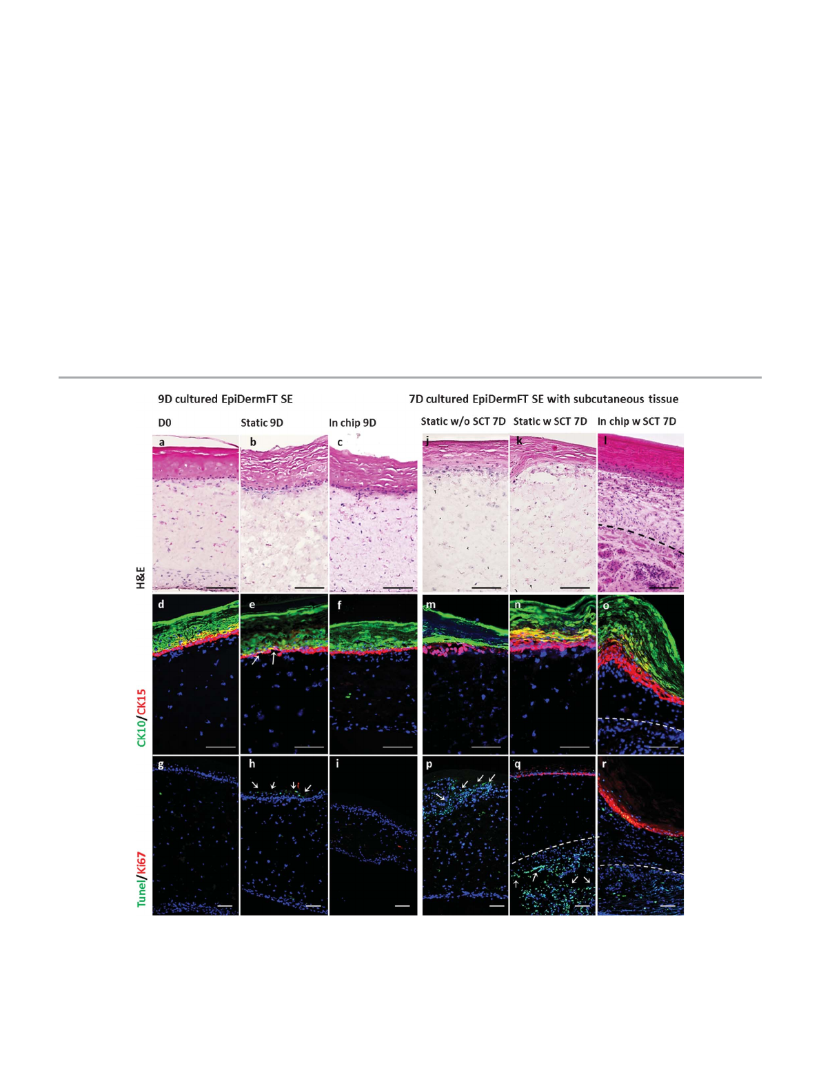
Fig. 2 In vitro skin equivalents (Mattek, Ashland, US) cultured for 9 days in MOC or static conditions in comparison to the same SE cultured for 7 days with
subcutaneous tissue (SCT) from prepuce. Haematoxylin and eosin (H&E) staining for histological comparison of the sections (a–c, j–l) and immunofluorescence
staining for epidermal markers Cytokeratin 10 and 15 (d–f, m–o) are applied. Overlapping markers are visualized as yellow. Finally, Ki67 and TUNEL assay for
proliferation and apoptosis (g–i, p–r) are used for comparison of viability of tissues (arrows indicate discontinuous basal layer of epidermis in e, TUNEL positive
apoptotic cells in p and disintegrated tissue in q). Dashed lines mark the border between SE and SCT. Scale bars indicate 100 mm for each picture.
This journal is ßThe Royal Society of Chemistry 2013 Lab Chip, 2013, 13, 3555–3561 |3557
Lab on a Chip Paper
Published on 01 May 2013. Downloaded by TU Berlin - Universitaetsbibl on 29/03/2016 10:22:02.
View Article Online
A Keyence BZ-9000 Microscope with the BZ-II-Viewer soft-
ware was used for all microscopic imaging and the images
were merged using the BZ-II-Analyser software (Keyence, Neu
Isenburg, Germany). Phase contrast and fluorescent images
were adjusted for tone levels, brightness and contrast.
Results and discussion
9-day cultured EpiDermFT skin equivalent
We prolonged the recommended culture period for static
conditions of EpiDermFT SE to a total culture period of 9 days.
We could observe a rearrangement and compression of the
dermal matrix structure in dynamic MOC cultures as cell
nuclei are located closer to each other with denser extracellular
matrix structure compared to day 0 and the control. There
were more viable cells in the SEs cultured in the MOC
compared to the SEs with static culture conditions (Fig. 2a–c).
The different layers of the epidermis were stained by
immunofluorescence showing the expression of the epidermal
markers CK10 for differentiating keratinocytes and CK15 for
undifferentiated keratinocytes. Both were similarly expressed
in static and dynamic MOC cultures. Although epidermal
barrier function seemed to be preserved better in the MOC
culture according to the continuous lining of the CK15 positive
cells at the basal layer of the epidermis, there is discontinuity
in static culture as indicated with arrows (Fig. 2d–f). Hardly
any proliferating and apoptotic cells were seen in the MOC and
day 0 control sections according to Ki67 TUNEL double
staining (Fig. 2g, i), whereas selected apoptotic cells could be
found in the epidermal layer in static cultures (Fig. 2h),
suggesting the onset of degradation of this SE.
7-day cultured EpiDermFT skin equivalent with sub-cutaneous
tissue
We used EpiDermFT
TM
SE in combination with SCT dissected
from a prepuce. SCT is composed mainly of adipocytes,
fibroblasts and macrophages. Considering the support of lipid
metabolism and paracrine effects of adipose-derived cells on
the upper levels of skin, we integrated SCT to EpiDermFT
TM
SE
under the same experimental conditions for 7 days. We
observed more viable cells and a compressed dermal matrix in
the samples with SCT cultured in the MOC compared to static
cultures with and without SCT stained by H&E (Fig. 2j–l). SCT
tissue was well integrated to SE in the MOC (Fig. 2l), while its
integration was poor in the static culture (Fig. 2q, arrows
indicating disintegrated tissue). CK15 positive cells are more
abundant in the MOC culture (Fig. 2o) showing the greatest
similarity to the native tissue (Fig. 3d as reference), even
though there is an increase of undifferentiated keratinocytes
in the static culture with SCT compared to regular static
culture (Fig. 2m, n). In contrast to static cultures, proliferating
cells are observed at the basal layer of the epidermis in
cultures with SCT and even more in the dynamic cultures
(Fig. 2p–r). SCT is expected to have a stimulating effect on
epidermal cells and this effect seemed to be even more
prevalent in the dynamic culture. Considering the absence of
proliferating cells in the epidermis in the cultures without
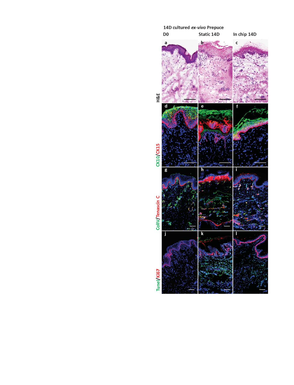
Fig. 3 Maintenance of ex vivo prepuce 14 days in culture. Day zero conditions of
the tissue are shown in a, d, g, and j. Static cultures are shown in b, e, h, and k,
while dynamically cultured tissue in MOC is indicated with c, f, i and l. H&E
staining is applied for histological comparison of the tissues (a–c).
Immunofluorescence staining for epidermal markers Cytokeratin 10 and 15 (d–
f), basement membrane markers Tenascin (TenC) and Collagen IV (ColIV) (g–i),
finally, Ki67 and TUNEL assay for proliferation and apoptosis (j–l) are used for
the evaluation of the viability of the tissues. Overlapping markers are visualized
as yellow. Scale bars indicate 100 mm for each picture.
3558 |Lab Chip, 2013, 13, 3555–3561 This journal is ßThe Royal Society of Chemistry 2013
Paper Lab on a Chip
Published on 01 May 2013. Downloaded by TU Berlin - Universitaetsbibl on 29/03/2016 10:22:02.
View Article Online
SCT, epidermal integrity is expected to be weakened (Fig. 2h,
p). A massive increase in the amount of apoptotic cells is seen
in the SCT of the static culture. The tissue itself is also poorly
integrated to the SE (Fig. 2q). Taking these results into
account, a higher metabolism of the ex vivo tissue seems to
require dynamic perfusion.
14-day cultured ex vivo prepuce
We evaluated SB maintenance in culture for future substance
testing in comparison to SEs. Histological evaluation by H&E
showed epidermal disruption and dermal reorganisation in
the static cultures (Fig. 3b), whereas MOC-cultured biopsies
showed similar distribution to the control tissues (Fig. 3a, c).
In addition, CK10/CK15 staining showed a disintegrated
epidermis in the static culture compared to day 0 and MOC
culture (Fig. 3d–f). ColIV and TenC, which are both synthe-
sised and secreted by keratinocytes and fibroblasts, were used
as markers for the extracellular basement membrane.
17
ColIV
is mainly found in the basement membrane of the skin,
including the epidermal-dermal border and the dermal-
endodermal border including blood vessels.
18
TenC is an
extracellular matrix component known to be important for cell
shape and migration behaviour. It has a pro-migratory role for
epidermal cells around the basement membrane in skin and
has been shown to be upregulated during wound healing,
inflammatory processes and fibrosis.
19,20
For native skin
expression pattern of ColIV and TenC, see Fig. 3g. ColIV
expression was generally preserved in contrast to TenC
(Fig. 3g–i). Static SB culture shows elevated levels of TenC
below the basement membrane, suggesting induced fibrotic
processes (Fig. 3h). Proliferation in the epidermis was
maintained in the MOC according to Ki67 positive cells, while
it was decreased in static culture compared to day 0. TUNEL
positive cells in dermis are more abundant in the static culture
compared to the MOC culture, suggesting a higher amount of
cell loss (Fig. 3j–l).
14-day cultured ex vivo follicular unit extracts
We used the MOC system to culture FUEs as a further step in
the attempt to emulate the biology of the skin and its
appendages. In contrast to the well-established Philpott assay
using single, truncated hair follicles to study hair follicle
biology in vitro,
21
we cultured complete hair follicular units.
These almost intact philosebaceous units include the perifol-
licular epidermis, dermis and the sebaceous gland(s). We
aimed to prolong the culture period of the ex vivo hair follicles,
taking into account the support of the glands and the
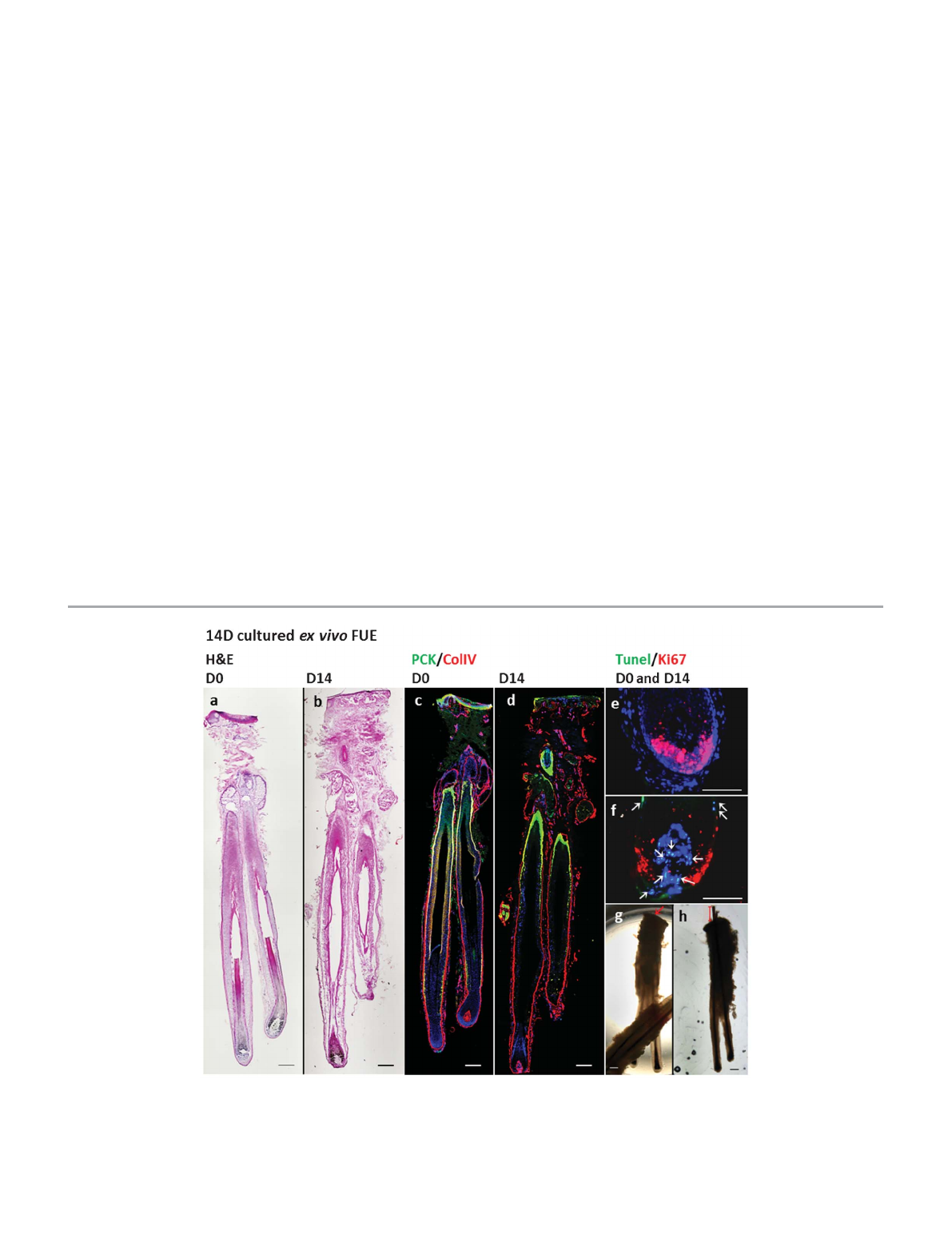
surrounding skin tissue on hair follicle maintenance. H&E
staining showed partial loss of structural integrity and a
decrease in the total number of cell nuclei within the central
and proximal hair follicle after 14 days of culture (Fig. 4a, b),
indicating signs of the ongoing regression phase (catagen) of
the hair follicle. Nevertheless, a pan cyto-keratin and ColIV
double immunostaining revealed an intact appearance of the
Fig. 4 FUEs cultured in the MOC for 14 days. H&E staining for day 0 and day 14 sample (a, b). Immunofluorescence staining for collective CKs (PCK) and ColIV for day 0
and day 14 samples (c, d) TUNEL assay and Ki67 staining shown for bulb region of the hair for day 0 and day 14 sample (e, f) (arrows indicate TUNEL positive cells in
the DP and CTS. Light microscopy images from day 0 and day 14 indicating hairs-shaft elongation in culture (g, h). Scale bars indicate 200 mm for a, b, c and d; 50 mm
for e and f; 300 mm for g and h.
This journal is ßThe Royal Society of Chemistry 2013 Lab Chip, 2013, 13, 3555–3561 |3559
Lab on a Chip Paper
Published on 01 May 2013. Downloaded by TU Berlin - Universitaetsbibl on 29/03/2016 10:22:02.
View Article Online
Loading more pages...