
A Threestepped Coordinated Level Set Segmentation Method for
Identifying Atherosclerotic Plaques on MR-Images
Oliver Gloger*, Matthias Ehrhardt**, Thore Dietrich***, Olaf Hellwich, Kristof Graf***,
Eike Nagel****
Berlin University of Technology (TUB), Computer Vision and Remote Sensing
Franklinstraße 28/29, Sekretariat FR3-1, 10587 Berlin, Germany
Tel.: ++49-30-314-73109 , Fax: ++49-30-314-21104,
berlin.de, [email protected], [email protected]
Abstract
In this work we propose an adapted level set segmentation technique for the recognition of
atherosclerotic plaque tissue on magnetic resonance images. The images are 2dimensional
cross-sectional images and show different profiles from ex-vivo human vessels with high
variability in vessel shape. We used a curvature based anisotropic diffusion technique to
denoise the magnetic resonance images.
The segmentation technique is subdivided into three level set steps. Hence, the result of every
phase serves as constructive knowledge for the next level set step. By analyzing and
combining carefully all available channel information during the first and second step we are
capable to delineate exactly the vessel walls by using and adapting two well-known level set
segmentation techniques.
The third step controls an enclosing level set which separates the plaque patterns from healthy
media tissue. In this step we introduce a local weighting concept to consider intensity
information for conspicuous plaque patterns. Furthermore, we propose the introduction of a
maximal shrinking distance for the third level set in the vessel wall and compare the results of
the local weighting algorithm with and without the concept of the maximal shrinking distance.
The incorporation of locally weighted intensity information into the level set method allows
the algorithm to automatically distinguish plaque from healthy media tissue. The knowledge
of the maximal shrinking distance can improve the segmentation results and enables to
delineate tissue areas where plaque is most likely.
Keywords: level set segmentation, active contours, medical image segmentation, anisotropic
diffusion, atherosclerotic plaques, Canny edges
* Chair for Computer Vision and Remote Sensing (TU Berlin) /
Ernst Moritz Arndt University Greifswald, Institute for Community Medicine, Walter-Rathenau-Str. 48,
17475 Greifswald
** Weierstrass Institute for Applied Analysis and Stochastics, Mohrenstr.39, 10117 Berlin, Germany
*** German Heart Center Berlin, Augustenburger Platz 1, 13353 Berlin
**** King's College London, St. Thomas’ Hospital, London SE1 7EH
1

1. Introduction and Motivation
Atherosclerosis is a serious disease affecting the arterial blood vessels. It is a continuous
process in which certain substances (e.g. Cholesterol) accumulate within the vessel wall. This
accumulation may cause a slow reduction of the vessel lumen causing a deterioration of blood
flow during exertion, or – in the case of a “vulnerable” plaque - rupture suddenly causing a
closure of the vessel, which leads to a myocardial infarction (heart attack) or stroke. Thus, it is
of most importance to discriminate between stable and unstable plaque and predict potential
plaque rupture.
With magnetic resonance (MR) imaging (MRI) it is possible to visualize all parts of the body
– including the heart and the vessels - with high spatial resolution (typical values for in-plane
resolutions in clinical research are about 400 µm x 400 µm for human plaques in coronary
arteries and about 500 µm x 500 µm for aorta plaques). It is possible to visualize plaque in
larger vessels, such as the aorta and the carotid arteries with state of the art MR scanners and
to characterize plaque components. Methods for automatic plaque segmentation of carotid
plaque have been proposed [1] and allow for serial assessment of plaque size and its lipid
core. Similarly vessel wall imaging of the coronary arteries is possible and first clinical data is
available [2, 3, 4]. However, the rapid motion of the coronary arteries in combination with the
small vessel size has made plaque characterization in the coronary arteries impossible.
Potential future developments will improve spatial resolution, overcome motion artifacts by
motion compensation algorithms [5, 6] or specifically enhance those components of a plaque
which cause plaque rupture using specific contrast agents [7, 8].
Due to prospective improvements of MR-image resolution we aim to test modern
segmentation techniques for higher in-plane resolutions of 79 µm x 79 µm, which is of course
five to six times higher than usual. Informations about segmentation success for this type of
MR-images should help to estimate the benefit of level set segmentation techniques for plaque
images in general. If we expect higher resolutions for aorta or coronary arteries in future, this
contribution represents a pioneer work for automatic segmentation of prospective image
resolutions.
Primarily, we applied pixel-oriented classification algorithms (i.e. k-means) and low-level
segmentation approaches (i.e. watershed-segmentation, region growing, presegmentation with
scale space concepts [9]) for plaque identification, but stated that suchlike approaches failed
in most circumstances. To facilitate automatic image analysis and to reduce observer
variability we propose a new algorithm for the assessment of plaque images. In contrast to
previous methods with probabilistic approaches we provide a technique that subdivides
healthy media tissue from plaque with a closed contour into two resulting subparts. This
bordering closed contour guarantees an important local closeness to distinguish between
plaque and healthy media tissue during the segmentation process.
Atherosclerotic plaques cannot be differentiated on MR-images by the information of gray
values alone, but have an associated regional context. We know however, that atherosclerotic
plaques are located within or adjacent to the arterial walls. Consequently, we are interested in
a segmentation technique that – in a first step- can segment the arterial vessels and then – in a
second step- can recognize relevant plaque structures in the vessel walls. Since vessel
boundaries in 2D cross-sections must present closed loops, we used a segmentation technique
based on active contours as an appropriate starting point.
2

The model of active contours has been introduced by Kass et. al. [10] and represents an
explicit form for describing the motion of a closed contour. The algorithm is based on the
principle, that a contour continues to move towards the boundaries of an object. This process
can be represented by a minimization of an explicit energy function which includes two
terms: one for controlling the smoothness of the contour and one to influence the attraction of
the contour to the object boundaries. This parametric model has been frequently used and
extended for segmentation problems in (bio)medical image segmentation [11, 12, 13, 14, 15,
16].
The classical model of snakes and active contours yields a significant disadvantage of
managing certain topological changes during the contour evolution process. Operations like
splitting and merging are not possible to represent with this classical approach. Unfortunately,
those flexible curve propagations could be of great importance during the segmentation
process of detailed medical shapes especially for the delineation of the outer media border and
the recognition of plaque (which we show in detail in chapter 4.2). Caselles et al. [17]
associated the classical active contour model with a geodesic flow model which is represented
by a partial differential equation based on a mean curvature flow. The energy minimizing
problem can be solved by finding the geodesic contour in Riemann space derived from the
image. Solving this model with a level set approach provides the contour with the ability of
managing the desired topological changes. The contour is now represented implicitly through
the level set equation and yields additionally the advantage of having fewer parameters to
guide the propagation of the contour. Parameters used in the level set propagation are
geometric measures which are independent of the parametrization of the curve. So they are
also called geometric active contours where the classical snakes refer to parametric active
contours.
In combination with variational techniques the level set method has often been applied for
finding the minima of energy functionals, which aim to solve special segmentation problems
for modern computer vision applications. In [18] a dynamical framework is proposed that
uses variational techniques derived from a Bayesian model and is able to segment scalar and
vector-valued images. In contrast to the edge based energy functionals [17], region based
energy functionals [19, 20] are independent of edge information making their application for
medical images very promising. However, variational formulations that use either edge based
terms or region based terms have the disadvantage that their derived level set propagation
stops in local minima. Recently, some approaches try to overcome those problems. In [21] a
local term is incorporated in a region based energy functional [19] that minimizes the local
contrast variances on a narrow band inside and outside of the contour. Lankton et al. [22]
combine the advantages of region based [19] and edge based energy functionals [17] by
deriving a geodesic energy from local regions around the contour which results in a hybrid
curve evolution.
The exclusive application of region based and edge based energy functionals is not sufficient
for atherosclerotic MR-images due to their vessel complexity that show high shape and
intensity variances and lacks of local edge information. In this work we propose a level set
technique that uses relevant informations during propagation to overcome those challenges
and is capable for plaque delineation on a variety of MR-images.
In the following Chapters we explain this new approach for plaque segmentation successively.
In Chapter 2 we describe the image data we work with and explain how they have been
created. In Chapter 3 we describe the denoising method we used for the MR-images as
preprocessing step in our algorithm. In Chapter 4.1 we give a short summary of the theoretical
3
aspects of the level set method which are crucial for understanding the subsequential
concepts. Chapter 4.2 describes our threestepped level set method in detail and shows the
results of the segmentation process of the inner and outer media borders. Furthermore, this
Chapter introduces the proposed concepts for the local weighting of conspicuous vessel wall
areas and the healthy media border. Chapter 5 illustrates and compares the results for the
proposed concepts. Finally, in Chapter 6 we give a summary and a conclusion of this work
and summarize our ideas for future work.
2. MR Data Acquisition
Human arterial specimens from the groin (iliac region) were obtained during vessel
replacement surgery. After freezing at -80°C, each specimen was warmed up to 30°C in a
phosphate buffered solution. Then it was placed in a 15 ml tube which was then filled with
1% agarose solution. The specimens were measured in a 7 Tesla MR-Tomograph (Bruker
Germany, PharmaScan). A whole body mouse coil (diameter of 38 mm) was utilized to obtain
a 79 µm x 79 µm in-plane resolution and a 110 µm slice thickness. Scan time for four
contrasts (PD, T1, T1fatsat, T2) was about 16 hours. The method is described in more detail
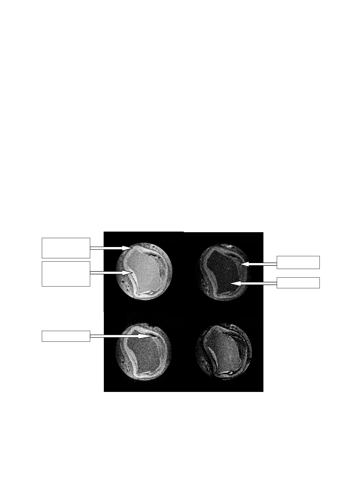
in [23]. Figure 1 shows an imaging example of 4 channels each with a 256 x 256 matrix size.
With those different channels we have the possibility to emphasize special tissue patterns in a
certain range.
PD
T1
muscular vessel
wall (media)
chronic plaque
young plaque
(neointima
formation) Lumen
T1fatsat
T2
calcification
Fig.1: MR-images of arterial profiles quantified with 4 different types of measurement (proton-density, T1, T1-
fatsaturated, T2). The important parts of the arterial profiles are inscribed to the images above.
4

3. Denoising of MR-images
Although certain relevant plaque patterns are well visible the images exhibit regions with a
low signal-to-noise ratio (SNR). The level set propagation is influenced by high image
gradients. To ensure a suitable level set propagation we have to denoise those images
effectively without loosing too much detail information. Blurring the images with a Gaussian
kernel causes an additional dislocation and blurring effect for important edges. Those edges
are indispensable information for the stopping decisions of the level set propagation.
Consequently, we decided to apply edge-preserving algorithms based on anisotropic
diffusion.
A frequently referenced non-linear diffusion model was introduced by Perona and Malik [24].
In [25, 26] two important extensions of the Perona-Malik model have been proposed. Wei
[25] suggests a generalisation of the Perona-Malik equation by introducing an edge enhancing
functional and using local statistics for more effective edge preservation and noise removal.
Another extension of the Perona-Malik model (with representing the underlying image) has
been developed by Whitaker and Xue [26], which they call a modified curvature diffusion
equation (MCDE):
u
(( ) )
uu
udivg u
tu
∂∇
=∇ ⋅ ∇ ⋅
∂∇
(1)
They propose it as a level-set analogue of the anisotropic diffusion model of Perona and
Malik and also emphasize its superiority compared to the Perona-Malik model. Their curve
evolution is based upon the principles of the self-snakes, which have been introduced by
Sapiro [27].
Due to the fact that we use denoising techniques as a preprocessing step for our work, we
chose the implementation of the ITK development framework [28] to denoise MR images
using the MCDE model. The diffusion process can be controlled by the number of iterations
and a conductance value, which influences the conductance term of the diffusion process. In
the ITK the conductance term [28] is used as a function that can suppress diffusion at higher
image gradients and preserve edges by
()
2
2
exp 2
u
gu k
⎛⎞
∇
∇
=⎜−
⎜⎟
⎝⎠
⎟
(2)
and can be steered by the conductance value k. We used 175 iterations for time steps of 0.125
seconds and denoised the images with conductance values in the range of 1.0 to 2.5.
5
Loading more pages...