972 https://doi.org/10.1107/S2056989019008053 Acta Cryst. (2019). E75, 972–975
research communications
Received 9 May 2019
Accepted 4 June 2019
Edited by O. Blacque, University of Zu
¨rich,
Switzerland
Keywords: crystal structure; coordination
compound; copper(II) complex; dinuclear
complex; bipyridine derivative; tartrates;
electroless copper baths; hydrogen bonding;
stacks.
CCDC reference:1920829
Supporting information:this article has
supporting information at journals.iucr.org/e
Synthesis, characterization, and crystal structure of
aquabis(4,4000-dimethoxy-2,2000-bipyridine)[l-(2R,3R)-
tartrato(4)]dicopper(II) octahydrate
Dennis Wiedemann,* Roman-David Kulko and Andreas Grohmann
Institut fu
¨r Chemie, Technische Universita
¨t Berlin, Strasse des 17. Juni 135, 10623 Berlin, Germany. *Correspondence
e-mail: [email protected]erlin.de
Typical electroless copper baths (ECBs), which are used to chemically deposit
copper on printed circuit boards, consist of an aqueous alkali hydroxide
solution, a copper(II) salt, formaldehyde as reducing agent, an l-(+)-tartrate as
complexing agent, and a 2,20-bipyridine derivative as stabilizer. Actual
speciation and reactivity are, however, largely unknown. Herein, we report on
the synthesis and crystal structure of aqua-1O-bis(4,40-dimethoxy-2,20-bipyri-
dine)-1
2
N,N0;2
2
N,N0-[-(2R,3R)-2,3-dioxidosuccinato-1
2
O
1
,O
2
:2
2
O
3
,O
4
]di-
copper(II) octahydrate, [Cu
2
(C
12
H
12
N
2
O
2
)
2
(C
4
H
2
O
6
)(H
2
O)]8H
2
O, from an
ECB mock-up. The title compound crystallizes in the Sohncke group P2
1
with
one chiral dinuclear complex and eight molecules of hydrate water in the
asymmetric unit. The expected retention of the tartrato ligand’s absolute
configuration was confirmed via determination of the absolute structure. The
complex molecules exhibit an ansa-like structure with two planar, nearly parallel
bipyridine ligands, each bound to a copper atom that is connected to the other
by a bridging tartrato ‘handle’. The complex and water molecules give rise to a
layered supramolecular structure dominated by alternating stacks and
hydrogen bonds. The understanding of structures ex situ is a first step on the way
to prolonged stability and improved coating behavior of ECBs.
1. Chemical context
The production of printed circuit boards (PCB) starts with
electroless copper deposition (ECD) on electrically non-
conductive plastics. Copper is deposited from an alkaline
solution of a copper(II) salt and a reducing agent (in general,
formaldehyde). The reduction of copper(II) ions proceeds
only at pH > 10, thus making methanediolate (deprotonated
formaldehyde hydrate) the actual reactant (Van Den Meer-
akker, 1981; Jusys & Vaskelis, 1992). A complexing agent
prevents the precipitation of copper(II) hydroxide (K
L
=
0.16 mmol
3
L
3
), which would otherwise occur at pH > 5.7.
Since the early development of ECD in 1946, l-(+)-tartrate
has commonly been used as complexing agent (Narcus, 1947).
Between pH 11 and 13, it forms bis(tartrato)copper(II),
[Cu(C
4
H
2
O
6
)
2
]
6–
, where each tartaric-acid-derived ligand is
quadruply deprotonated. This complex is also known from
Fehling’s solution (Fehling, 1848; Ho
¨rner & Klu
¨fers, 2016).
Reactant solutions facilitating ECD, so-called electroless
copper baths (ECB), are metastable with respect to the
precipitation of metallic copper, making additional stabilizers
necessary. Over the past 60 years, a plethora of compounds has
been used for this purpose (Agens, 1960; Saubestre, 1972),
affecting not only the lifetime of ECBs but also the rate of
ISSN 2056-9890
ECD and the physical properties of the deposited copper.
Amongst the stabilizers, 2,20-bipyridine and its derivatives are
especially popular (Oita et al., 1997).
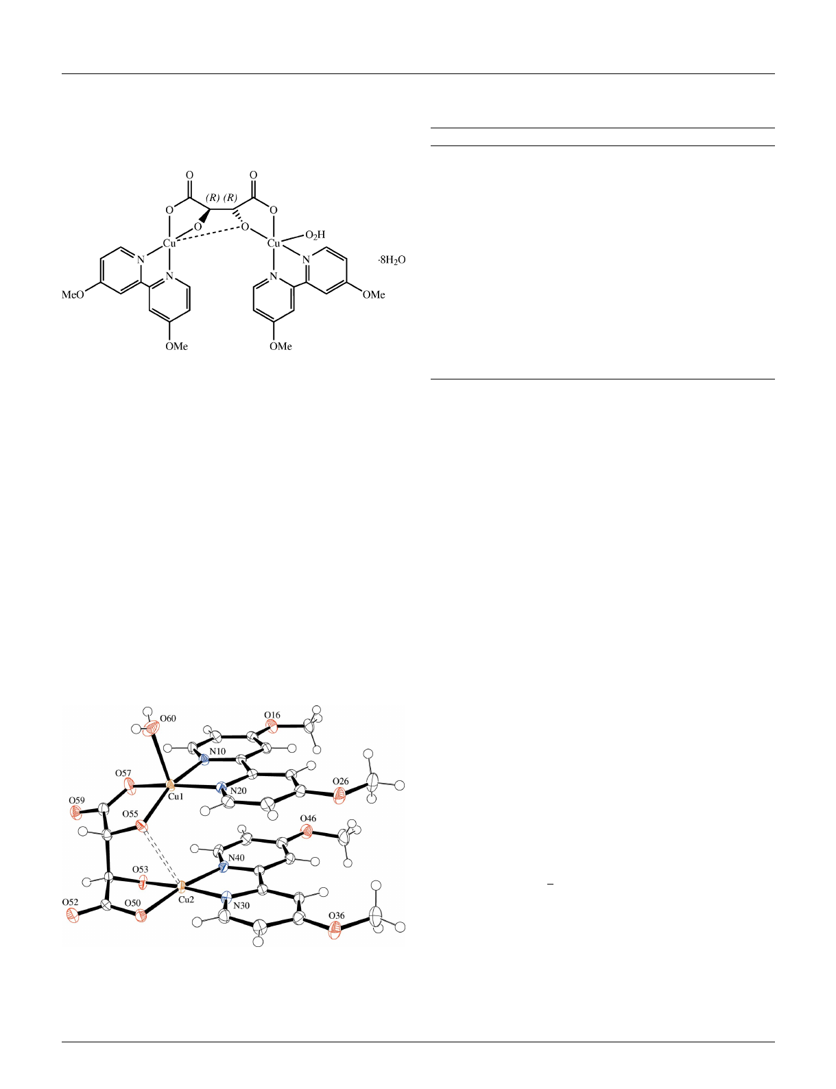
Herein, we report on the crystal structure of a compound
that formed from an alkaline solution of a copper(II) salt, a
tartrate, and 4,40-dimethoxy-2,20-bipyridine (dmobpy) during
the investigation of stabilities of various copper(II) complexes
with ligands derived from 2,20-bipyridine (bpy).
2. Structural commentary
The compound crystallizes in the Sohncke group P2
1
with one
chiral complex molecule and eight molecules of hydration
water in the asymmetric unit. The copper(II) ions in the
dinuclear complex (see Fig. 1) are each coordinated by two
azine nitrogen donors, one alcoholate and one carboxylate
oxygen donor. The lengths of the respective short bonds (ca
1.89–2.00 A
˚) reflect the formal charge of the donor atoms,
while a cis configuration is enforced by the structure of the
ligand. An additional longer bond to an aqua ligand [d(Cu1—
O60) = 2.322 (3) A
˚] augments the coordination environment
of Cu1 to a distorted square pyramid. Cu2, on the other hand,
is coordinated in a square planar fashion with a short contact
to a second alcoholate oxygen atom [d(Cu2O55) =
2.549 (2) A
˚].
The 4,40-dimethoxy-2,20-bipyridine ligands are nearly planar
[positional root-mean-square (r.m.s.) deviation excluding
hydrogen atoms: 0.032 A
˚for ligand containing N10 and N20,
0.041 A
˚for ligand containing N30 and N40], almost parallel
[interplanar angle: 2.70 (4)], and give rise to intramolecular
stacks with an average centroid–plane distance of 3.36 (5) A
˚.
Because of this, the overall molecular structure resembles that
of ansa compounds, with the tartrato ligand representing the
‘handle’. The tartrato ligand assumes an antiperiplanar (ap)
conformation with respect to the central bond of the carbon-
atom chain. The C—O bonds at the carboxylate donors are
synperiplanar (sp) to the C—O bonds at the neighboring
alcoholate donors.
The absolute structure of the crystal was established via
anomalous-dispersion effects [the inversion-distinguishing
power of the experiment is strong according to Flack &
Bernardinelli 2000)] and matches the absolute configuration
of the employed l-(+)-(2R,3R)-tartrate. The Flack parameter
is within the statistical range for an untwinned crystal, thus
confirming the enantiopurity of the complex molecules (Flack
& Bernardinelli, 2000).
3. Supramolecular features
Roughly parallel to {111}, complexes form infinite stacks, in
which the intermolecular distance of 3.37 (6) A
˚(average
centroid–plane distance) equals the intramolecular one (see
Fig. 2a). A hydrogen bond from the aqua ligand to the
carboxylato oxygen atom O50 of the neighboring molecule in
the stack connects the tartrato(4) ligands, forming an infinite
hydrophilic backbone along the adirection.
The eight unique water molecules constitute a local network
of hydrogen bonds (see Table 1) in a pocket formed by aqua
research communications
Acta Cryst. (2019). E75, 972–975 Wiedemann et al. [Cu
2
(C
12
H
12
N
2
O
2
)
2
(C
4
H
2
O
6
)(H
2
O)]8H
2
O973
Table 1
Hydrogen-bond geometry (A
˚,).
D—HAD—H HADAD—HA
O60—H60AO50
i
0.81 (5) 2.00 (5) 2.802 (3) 167 (5)
O60—H60BO67 0.77 (5) 1.98 (5) 2.750 (4) 177 (5)
O61—H61AO65 0.81 (2) 2.02 (3) 2.813 (4) 166 (4)
O61—H61BO52
ii
0.82 (2) 1.92 (2) 2.739 (3) 177 (4)
O62—H62AO64 0.79 (2) 2.01 (3) 2.774 (3) 164 (5)
O62—H62BO55 0.80 (2) 1.85 (2) 2.650 (3) 174 (5)
O63—H63AO59
ii
0.83 (2) 1.97 (2) 2.806 (3) 178 (4)
O63—H63BO61 0.85 (2) 2.13 (2) 2.970 (3) 176 (4)
O64—H64AO52 0.81 (2) 2.05 (3) 2.762 (3) 146 (4)
O64—H64BO65 0.85 (2) 1.95 (3) 2.719 (3) 149 (4)
O65—H65AO59
iii
0.82 (2) 2.08 (3) 2.871 (3) 162 (4)
O65—H65BO66 0.81 (2) 2.01 (2) 2.801 (4) 167 (4)
O66—H66AO53
ii
0.79 (2) 1.91 (3) 2.680 (3) 169 (5)
O66—H66BO62 0.80 (2) 2.02 (3) 2.808 (4) 167 (5)
O67—H67AO62 0.84 (2) 1.91 (3) 2.737 (4) 168 (5)
O67—H67BO59
ii
0.83 (2) 2.15 (2) 2.973 (3) 178 (5)
O68—H68AO57
iii
0.90 (3) 2.52 (4) 3.236 (4) 138 (5)
O68—H68BO60
iv
0.91 (2) 2.23 (3) 3.129 (5) 169 (5)
Symmetry codes: (i) xþ1;y;z; (ii) x;y;z1; (iii) x1;y;z1; (iv) x1;y;z.
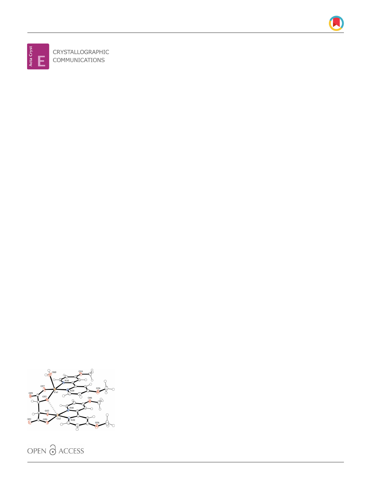
Figure 1
Molecular structure of the title compound as an ORTEP plot (complex
molecule only, solvent water molecules omitted for clarity). Hydrogen
atoms are depicted as spheres with arbitrary radius, all other atoms as
displacement ellipsoids of 50% probability. The dashed line indicates a
non-bonding short contact.
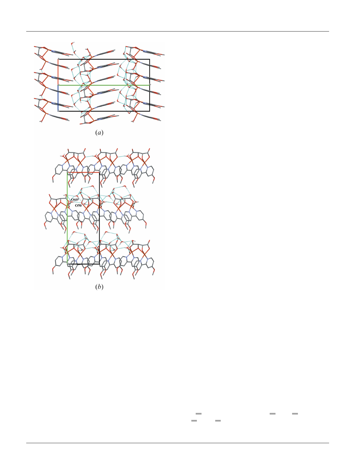
(donors only) and tartrato ligands (all oxygen atoms as
acceptors). The methoxy groups do not partake in hydrogen
bonding but build a hydrophobic lining of the pocket. In this
way, a front-to-back arrangement of alternating water and
complex layers along bis formed (see Fig. 2b).
4. Database survey
The Cambridge Structural Database [CDS 5.40 Update 1
(February 2019); Allen, 2002; Groom et al., 2016] contains 33
structures of tartratometal (Co
II
,Cr
III
,Cu
II
,Pd
II
,Pt
II
)
complexes with bipyridine-related ligands, amongst which
twelve contain copper(II). The palladium(II) and platinum(II)
complexes are structurally loosely related to
[Cu
2
(dmobpy)
2
(-C
4
H
2
O
6
)(H
2
O)] in that they form isolated
neutral dinuclear complexes [{M
II
L}
2
(-C
4
H
2
O
6
)] (M: metal,
L: bipyridine-related ligand). Their centers, however, are
coordinated in a square-planar fashion without additional
longer bonds to oxygen donors.
The copper(II) complexes fall into two groups containing
either regular tartrate(2) or deprotonated tartrate(4). The
former group comprises isolated cationic complexes such as
[{Cu(bpy)
2
}
2
(-C
4
H
4
O
6
)]
2+
(Wu et al., 2008) and poly-/oligo-
meric complexes such as [Cu(bpy)(-C
4
H
4
O
6
)]
n
(Liu et al.,
2008). The latter group, on the other hand, incorporates
isolated neutral complexes like aqua-terminated [Cu
2
L
2
(-
C
4
H
2
O
6
)(H
2
O)] (L: bis[2-pyridyl]amine; Li et al., 2006) or
polymeric complexes bridged by carboxylate-Odonors such as
[{Cu(bpy)}
2
(
4
-C
4
H
2
O
6
)]
n
presenting Cu
2
O
2
motifs (Li et al.,
2005).
The closest known relative to the title compound, however,
is [Cu
2
(phen)
2
(-C
4
H
2
O
6
)(H
2
O)]8H
2
O (phen: 1,10-phenan-
throline), which crystallizes in the same space-group type with
comparable cell dimensions (Saha et al., 2011). Both structures
are crystal-chemically homeotypic and differ mainly in the
replacement of the 4,40-methoxy groups at the bipyridine-like
ligands by a 3,30-(1,2-ethenediyl) bridge.
5. Synthesis and crystallization
Copper(II) sulfate pentahydrate (4.96 g, 19.9 mmol, 1.00 eq),
potassium sodium l-(+)-tartrate tetrahydrate (12.33 g,
43.7 mmol, 2.20 eq), and sodium hydroxide (5.60 g,
140.0 mmol, 7.04 eq) were dissolved in deionized water (1 L),
resulting in a solution with pH = 12.8. 4,40-Dimethoxy-2,20-
bipyridine (216 mg, 1.00 mmol) was dissolved in sulfuric acid
(10 mL, 0.1 mol L
1
). In a plastic centrifuge tube, the tartra-
tocopper solution (5 mL) was mixed with the bipyridine
solution (0.12 mmol). The mixture was then filled up to a final
volume of 7 mL with deionized water and sodium hydroxide
solution to adjust the final pH to 12.8.
After two days of standing unsealed at ambient tempera-
ture, dark-blue crystals of [Cu
2
(dmobpy)
2
(-C
4
H
2
O
6
)(H
2
O)]-
8H
2
O formed.
An infrared (IR) spectrum in attenuated total reflectance
(ATR) was acquired from a ground crystal using a Thermo
Nicolet iS5 equipped with a Thermo Nicolet iD5 ZnSe sample
holder. Bands (vs: very strong, s: strong, m: medium, w: weak,
br: broad) were assigned using literature data (Hesse et al.,
1979; Socrates, 2001), as well as reference spectra of the
dmobpy ligand and potassium sodium l-(+)-tartrate. The
crystals were insoluble in common laboratory solvents
(alkanes, ethers, alcohols, dimethylformamide, dimethyl sulf-
oxide, and water) at ambient and elevated temperature and
decomposed in boiling coordinating solvents. Therefore, we
cannot provide data of analyses relying on solutions.
IR (ATR): ˜
= 3467, 3295 (all br w,[OH]), 1669 (s,
[OC O]), 1600 (vs,[OC—O], [C C], [C N]), 1558 (vs,
[C C], [C N]), 1499 (s), 1476, 1461, 1437, 1418 (all s,
[CH], dmobpy), 1344 (s,[CH], tartrato), 1317 (m), 1280 (vs,
s
[C—OMe]), 1253 (s, tartrato), 1226 (s, dmobpy), 1186 (w),
974 Wiedemann et al. [Cu
2
(C
12
H
12
N
2
O
2
)
2
(C
4
H
2
O
6
)(H
2
O)]8H
2
OActa Cryst. (2019). E75, 972–975
research communications
Figure 2
Packing diagram in views along (a) the vector a+cshowing the -stacked
bipyridine-type ligands and (b) the cell vector cshowing the layer-like
arrangement. Dashed bright blue lines indicate hydrogen bonds. Unit-cell
boundaries are depicted in black or red/green/blue marking the directions
a/b/c, respectively. Symmetry code: (i) x1, y,z.

1137 (w), 1103 (w), 1040, 1025, 1016, 1005 (all s,
as
[C—OMe],
[C C], [C N]), 872 (w), 851 (s,[CH]), 838 (vs,[CH]),
794 (s,
s
[COO]), 662 (br m,![COO]), 572 cm
1
(s,[COO]).
6. Refinement
Crystal data, data collection and structure refinement details
are summarized in Table 2. All non-hydrogen atoms were
refined with anisotropic displacement parameters. Hydrogen
atoms were located in difference-Fourier maps (for the
complex and most water molecules) or their positions were
inferred from neighboring sites (for the water molecule
containing O68). Carbon-bound hydrogen atoms were refined
with standard riding models. Oxygen-bound hydrogen atoms
were refined semi-freely with restrained 1,2- [d(O—H) ’
0.84 (2) A
˚] and 1,3-distances [d(HH) ’1.33 (4) A
˚], as well
as constrained isotropic displacement parameters [U
iso
(H) =
1.2U
eq
(O)]. Final bond lengths ranged between 0.77 (5) and
0.91 (2) A
˚with an r.m.s. deviation of 0.036 A
˚from the target
value.
After close inspection of the reflection statistics, data with
2>60
(essentially noise) and the high-angle reflection 1 21 0
(mismeasurement) were excluded from the final refinement.
The somewhat lower Friedel pair coverage is due to an
inadequate choice of data-collection strategy. Unfortunately,
we could not repeat the experiment because of sample loss.
Acknowledgements
We thank Ms Paula Nixdorf for the collection of diffraction
data and Dr. Julia Kohl (both Technische Universita
¨t Berlin)
for solubility assessment and spectroscopic analyses. We
acknowledge support by the German Research Foundation
and the Open Access Publication Fund of TU Berlin.
References
Agens, M. C. (1960). US-American Patent No. 2938805.
Allen, F. H. (2002). Acta Cryst. B58, 380–388.
Dolomanov, O. V., Bourhis, L. J., Gildea, R. J., Howard, J. A. K. &
Puschmann, H. (2009). J. Appl. Cryst. 42, 339–341.
Farrugia, L. J. (2012). J. Appl. Cryst. 45, 849–854.
Fehling, H. (1848). Arch. Physiol. Heilkd. 7, 64–73.
Flack, H. D. & Bernardinelli, G. (2000). J. Appl. Cryst. 33, 1143–
1148.
Groom, C. R., Bruno, I. J., Lightfoot, M. P. & Ward, S. C. (2016). Acta
Cryst. B72, 171–179.
Hesse, M., Meier, H. & Zeeh, B. (1979). Spektroskopische Methoden
in der organischen Chemie, pp. 55–92. Suttgart: Thieme.
Ho
¨rner, T. G. & Klu
¨fers, P. (2016). Eur. J. Inorg. Chem. pp. 1798–1807.
Jusys, Z. & Vaskelis, A. (1992). Langmuir,8, 1230–1231.
Li, D.-S., Wang, Y.-Y., Liu, P., Luan, X.-J., Zhou, C.-H. & Shi, Q.-Z.
(2005). Acta Chim. Sinica,63, 1633–1637.
Li, D.-S., Zhou, C.-H., Wang, Y.-Y., Fu, F., Wu, Y.-P., Qi, G.-C. & Shi,
Q.-Z. (2006). Chin. J. Chem. 24, 1352–1358.
Liu, J.-Q., Wang, Y.-Y., Ma, L.-F., Zhang, W.-H., Zeng, X.-R., Shi,
Q.-Z. & Peng, S.-M. (2008). Inorg. Chim. Acta,361, 2327–2334.
Macrae, C. F., Bruno, I. J., Chisholm, J. A., Edgington, P. R., McCabe,
P., Pidcock, E., Rodriguez-Monge, L., Taylor, R., van de Streek, J. &
Wood, P. A. (2008). J. Appl. Cryst. 41, 466–470.
Narcus, H. (1947). Met. Finish. 45, 64–70.
Oita, M., Matsuoka, M. & Iwakura, C. (1997). Electrochim. Acta,42,
1435–1440.
Parsons, S., Flack, H. D. & Wagner, T. (2013). Acta Cryst. B69, 249–
259.
Rigaku OD (2015). CrysAlis PRO. Rigaku Oxford Diffraction,
Yarnton, England.
Saha, R., Biswas, S. & Mostafa, G. (2011). CrystEngComm,13, 1018–
1028.
Saubestre, E. B. (1972). Plating 59, 563–566.
Sheldrick, G. M. (2015a). Acta Cryst. A71, 3–8.
Sheldrick, G. M. (2015b). Acta Cryst. C71, 3–8.
Socrates, G. (2001). Infrared and Raman Characteristic Group
Frequencies, 3rd ed. Chichester: Wiley.
Van Den Meerakker, J. E. A. M. (1981). J. Appl. Electrochem. 11,
387–393.
Westrip, S. P. (2010). J. Appl. Cryst. 43, 920–925.
Wu, Y.-P., Fu, F., Li, D.-S., Yang, Z.-H., Zou, K. & Wang, Y.-Y. (2008).
Inorg. Chem. Commun. 11, 621–625.
research communications
Acta Cryst. (2019). E75, 972–975 Wiedemann et al. [Cu
2
(C
12
H
12
N
2
O
2
)
2
(C
4
H
2
O
6
)(H
2
O)]8H
2
O975
Table 2
Experimental details.
Crystal data
Chemical formula [Cu
2
(C
12
H
12
N
2
O
2
)
2
(C
4
H
2
O
6
)-
(H
2
O)]8H
2
O
M
r
867.75
Crystal system, space group Monoclinic, P2
1
Temperature (K) 150
a,b,c(A
˚) 8.5134 (4), 23.7812 (9), 8.9028 (4)
() 101.401 (4)
V(A
˚
3
) 1766.87 (13)
Z2
Radiation type Mo K
(mm
1
) 1.29
Crystal size (mm) 0.84 0.70 0.09
Data collection
Diffractometer Agilent Xcalibur
Absorption correction Analytical (CrysAlis PRO; Rigaku
OD, 2015)
T
min
,T
max
0.440, 0.886
No. of measured, independent and
observed [I>2(I)] reflections
19893, 8971, 8476
R
int
0.021
(sin /)
max
(A
˚
1
) 0.703
Refinement
R[F
2
>2(F
2
)], wR(F
2
), S0.028, 0.072, 1.04
No. of reflections 8971
No. of parameters 536
No. of restraints 25
H-atom treatment H-atom parameters constrained
for H on C, refined H-atom
coordinates only for H on
heteroatoms
max
,
min
(e A
˚
3
) 0.49, 0.50
Absolute structure Flack xdetermined using 2429
quotients [(I
+
)(I
)]/[(I
+
)+(I
)]
(Parsons et al., 2013)
Absolute structure parameter 0.010 (6)
Computer programs: CrysAlis PRO (Rigaku OD, 2015), SHELXT (Sheldrick, 2015a),
SHELXL (Sheldrick, 2015b), ORTEP-3 for Windows (Farrugia, 2012), Mercury (Macrae
et al., 2008), OLEX2 (Dolomanov et al., 2009) and publCIF (Westrip, 2010).

supporting information
sup-1
Acta Cryst. (2019). E75, 972-975
supporting information
Acta Cryst. (2019). E75, 972-975 [https://doi.org/10.1107/S2056989019008053]
Synthesis, characterization, and crystal structure of aquabis(4,4′-dimeth-
oxy-2,2′-bipyridine)[µ-(2R,3R)-tartrato(4−)]dicopper(II) octahydrate
Dennis Wiedemann, Roman-David Kulko and Andreas Grohmann
Computing details
Data collection: CrysAlis PRO (Rigaku OD, 2015); cell refinement: CrysAlis PRO (Rigaku OD, 2015); data reduction:
CrysAlis PRO (Rigaku OD, 2015); program(s) used to solve structure: SHELXT (Sheldrick, 2015a); program(s) used to
refine structure: SHELXL (Sheldrick, 2015b); molecular graphics: ORTEP-3 for Windows (Farrugia, 2012) and Mercury
(Macrae et al., 2008); software used to prepare material for publication: OLEX2 (Dolomanov et al., 2009) and publCIF
(Westrip, 2010).
Aqua-1κO-bis(4,4′-dimethoxy-2,2′-bipyridine)-1κ2N,N′;2κ2N,N′-[µ-(2R,3R)-2,3-
dioxidosuccinato-1κ2O1,O2:2κ2O3,O4]dicopper(II) octahydrate
Crystal data
[Cu2(C12H12N2O2)2(C4H2O6)(H2O)]·8H2O
Mr = 867.75
Monoclinic, P21
a = 8.5134 (4) Å
b = 23.7812 (9) Å
c = 8.9028 (4) Å
β = 101.401 (4)°
V = 1766.87 (13) Å3
Z = 2
F(000) = 900
Dx = 1.631 Mg m−3
Mo Kα radiation, λ = 0.71073 Å
Cell parameters from 8483 reflections
θ = 3.7–32.5°
µ = 1.29 mm−1
T = 150 K
Plate, dark blue
0.84 × 0.70 × 0.09 mm
Data collection
Agilent Xcalibur
diffractometer
Radiation source: fine-focus sealed tube,
Agilent Enhance
Graphite monochromator
Detector resolution: 16.3031 pixels mm-1
ω scans
Absorption correction: analytical
(CrysAlis PRO; Rigaku OD, 2015)
Tmin = 0.440, Tmax = 0.886
19893 measured reflections
8971 independent reflections
8476 reflections with I > 2σ(I)
Rint = 0.021
θmax = 30.0°, θmin = 3.5°
h = −11→11
k = −33→31
l = −11→12
Refinement
Refinement on F2
Least-squares matrix: full
R[F2 > 2σ(F2)] = 0.028
wR(F2) = 0.072
S = 1.04
8971 reflections
536 parameters
25 restraints
Primary atom site location: dual
Secondary atom site location: difference Fourier
map
Hydrogen site location: mixed
Loading more pages...