
materials
Article
Nanoparticle Enhanced Antibody and DNA
Biosensors for Sensitive Detection of Salmonella
Sumeyra Savas 1, Aylin Ersoy 1, Yakup Gulmez 1, Selcuk Kilic 2, Belkis Levent 2and
Zeynep Altintas 3,*
1
National Research Institute of Electronics and Cryptology, The Scientific and Technological Research Council
2Turkey Public Health General Headquarter, Ankara 06100, Turkey; [email protected] (S.K.);
[email protected] (B.L.)
3Institute of Chemistry, Technical University of Berlin, Straße des 17. Juni 124, Berlin 10623, Germany
*Correspondence: [email protected]; Tel.: +49-30-314-23727
Received: 12 August 2018; Accepted: 22 August 2018; Published: 27 August 2018
Abstract:
Bacteria-related pathogenic diseases are one of the major health problems throughout
the world. Salmonella is a genus of rod-shaped Gram-negative enterobacteria of which more than
2600 serotypes have been identified. Infection with Salmonella can cause salmonellosis, a serious
bacterial toxi-infection syndrome associated with gastroenteritis, and paralyphoid and typhoid
fevers. Its rapid and sensitive detection is a key to the prevention of problems related to health.
This paper describes the development of antibody and DNA sensors for Salmonella detection using
a microfluidic-based electrochemical system. Commercial Salmonella typhimurium and Salmonella
typhimurium from human stool samples were investigated using standard and nanomaterial-amplified
antibody sensors. S. typhimurium could be detected down to 1 cfu mL
−1
. The specificity of
immunoassay was tested by studying with non-specific bacteria including E. coli and S. aureus
that revealed only 2.01% and 2.66% binding when compared to the target bacterium. On the other
hand, the quantification of Salmonella DNA was investigated in a concentration range of 0.002–200
µ
M
using the developed DNA biosensor that demonstrated very high specificity and sensitivity with
a detection limit of 0.94 nM. Our custom-designed microfluidic sensor offers rapid, highly sensitive,
and specific diagnostic assay approaches for pathogen detection.
Keywords:
Salmonella spp.; pathogen detection; antibody biosensor; DNA biosensor; microfluidic-
based electrochemical sensor; nanoparticle enhanced bio-detection; infectious diseases
1. Introduction
Foodborne diseases lead to diverse health problems worldwide [
1
,
2
]. The World Health
Organization reported that Salmonella typhimurium and Salmonella enteritidis are the most common
causes of foodborne illnesses all over the world. According to the reports of the European Food Safety
Authority, human salmonellosis has resulted in three billion euros loss per year [
3
]. In some regions,
more than 90% of Salmonella strains isolated from humans until 1970 was S. typhimurium, but now
incidence of S. enteritidis is also gradually increasing, which has been the most frequently isolated
serotype in the last 10 years [
4
]. The symptoms of Salmonella infection include abdominal pain, fever,
nausea, vomiting, diarrhea, dehydration, weakness, and loss of appetite, and the symptoms normally
appear 12–72 h after ingestion of contaminated foods or beverages [5].
There are several methods used for the detection of Salmonella serotypes, such as cultivation
techniques [
6
], enzyme-linked immunosorbent assays (ELISAs) [
7
], polymerase chain reaction (PCR)
Materials 2018,11, 1541; doi:10.3390/ma11091541 www.mdpi.com/journal/materials

Materials 2018,11, 1541 2 of 17
methods [
8
–
10
], and biosensors [
11
]. The gold standard for detection of Salmonella serotypes are
still conventional methods that are sensitive and inexpensive. However, these techniques require
more than five days to obtain a result and often lack in providing good specificity and sensitivity.
Additionally, the cultivation techniques are generally time consuming and the limit of detection for the
analysis is insufficient. The use of biosensor technology is a strong alternative to the other techniques
by offering highly sensitive, rapid, and easy-to-use bio-detection principles [
12
]. Today, biosensors
are widely used for pathogen detection and are able to measure bacteria down to 1 cfu mL
−1
. This is
particularly due to the significant impact of nanomaterials on the advancement of biosensors and
biosensing principles [
13
–
15
]. Furthermore, microbial biosensors often require sample volumes in the
microliter range and very short analysis time.
Various transducer systems based on surface plasmon resonance (SPR) [
16
], quartz crystal
microbalance (QCM) [
17
], and electrochemical sensing strategies [
18
] have been successfully employed
for bacteria detection. Offering very high sensitivity and good detection capacity electrochemical
sensors are among the widely used systems for Salmonella quantification [
14
,
19
,
20
]. Zhu et al.
developed a multichannel electrochemical immunosensor for Salmonella detection by combining the
rolling circle amplification with DNA-gold nanoparticles (AuNPs) probe [
20
]. Afonso et al. reported
a disposable immunosensor for electrochemical detection of Salmonella enterica subsp. Enterica serovar
Typhimurium LT2 using gold nanoparticles and magneto-immunoassay [
14
]. Sensitive amperometric
detection of Salmonella was also reported [
21
–
24
]. Even though great progress has been demonstrated
in the Salmonella detection, employing only one antibody in the biosensor design often results in
insufficient sensitivity, when the sensor could hardly discriminate between two LPS samples from two
different Gram-negative bacteria [25].
To overcome the selectivity problem, herein, we report antibody- and DNA-based biosensors
for Salmonella detection using a fully-automated custom-designed microfluidic sensing device
(MiSens) [
18
] that is composed of an electromechanical unit controlling the assay protocol via its
integrated software (MiCont
TM
). Normal and gold-nanoparticle amplified sandwich assays were
developed and used for the detection of commercial Salmonella samples and real samples from human
stool. DNA biosensor was developed by capturing the surface DNA probe on the neutravidin (NA)
immobilized sensor surface and then measuring the target Salmonella DNA based on the hybridization
reaction that occurs between the target DNA and the surface probe. As the measurement system relies
on the enzymatic reaction between horseradish peroxidase (HRP) and 3,3
0
,5,5
0
-tetramethylbenzidine
(TMB), the detector antibody and the DNA detection probe were both labeled with HRP. We have
demonstrated that the developed antibody and DNA biosensors are capable of measuring trace
amounts of Salmonella and Salmonella DNA, respectively.
2. Materials and Methods
2.1. Materials and Reagents
A monoclonal anti-Salmonella antibody was bought from BIO-RAD (Puchheim, Germany).
Peroxidase-labeled goat anti-Salmonella secondary antibody (BacTrace
®
Anti-Salmonella CSA-1 Antibody)
and Salmonella typhimurium were purchased from SeraCare Life Sciences (Gaithersburg, MD, USA).
Staphylococcus aureus and E. coli from human stool samples were obtained from the Public Health Agency
(Ankara, Turkey) for cross-reactivity studies. 11-Mercaptoundecanoic acid (MUDA), phosphate-buffered
saline tablets (PBS, 0.01 M phosphate buffer, 0.0027 M potassium chloride and 0.137 M sodium
chloride, pH 7.4), N-hydroxysuccinimide (NHS), analytical grade ethanol, horseradish peroxidase
(HRP), ethanolamine, and 3,3
0
,5,5
0
-tetramethylbenzidine (TMB) ready to use reagent with H
2
O
2
,
were purchased from Sigma Aldrich (Poole, UK). The gold nanoparticles in 15 nm (for DNA
assays) and 40 nm (for antibody assays) sizes were purchased from BBI International (Cardiff, UK).
Ultrapure water (18 M
Ω
cm
−1
) produced by a Milli-Q water system was used for analyses (Millipore Corp.,
Tokyo, Japan). 1-Ethyl-3-(3-dimethylaminopropyl)-carbodiimide (EDC) and biotin were purchased
Materials 2018,11, 1541 3 of 17
from Thermo Fisher Scientific (Loughborough, UK). The oligonucleotide sequences (target sequence:
5
0
-ACCGACGGCGAGACCGACTTT-3
0
; surface probe: Biotin-5
0
-CTCACCAGGAGATTACAACATGG;
detection probe: Biotin-3
0
-AGTGGCTAAAAGTCGGTCTC; control surface probe: Biotin-5
0
-
CAATATTTGGCGTGAATGGGTCGGAAAACA) for DNA sensor development were obtained from
Sentromer DNA Technologies LLC (Istanbul, Turkey).
2.2. Isolation of Salmonella Typhimurium from Human Stool Samples
Human stool sample was inoculated onto Salmonella-Shigella and xylose lysine deoxycholate agar
mediums, and incubated at 37
◦
C overnight. Bacterial culture was identified by using standard
biochemical tests (the use of glucose, citrate, and indole by bacteria, the production of gas and H
2
S
upon lactose fermentation of bacteria, the determination of the mobility and the ability to split urea).
The cultured colonies used glucose, citrate and indole confirmed the presence of Salmonella bacteria.
The isolate was serogrouped and serotyped using polyvalent and monovalent Salmonella antisera (Statens
Serum Institut, Copenhagen, Denmark) according to the Kauffmann-White scheme [
26
]. The colonies that
produced H
2
S were considered as a pure colony based on their morphology. After the confirmation and
serotyping, the strain was immediately frozen in tryptic soy broth with 16% glycerol at −80 ◦C.
2.3. Fully-Automated Microfluidic-Based Electrochemical Sensor with a New Chip Design
A custom-designed fully-automated electrochemical sensor was used in this study to develop
antibody- and DNA-based biosensors for Salmonella detection. The sensor fabrication and its developmental
stages were fully described in our earlier studies [
18
,
27
,
28
]. Meanwhile, we have realized several drawbacks
dealing with device’s electronics and mechanics. One of the main problems was electromagnetic noise
in the signal. In order to decrease the noise level, two modifications were employed: (1) Increasing of
the distance between the microfluidic pumps and the potentiometer. The microfluidic pump spread
unwanted magnetic pulses by the pulse width modulation (PWM) mechanism; (2) Applying a coaxial
cable structure instead of unscreened cable structure between the biosensor and potentiostat circuit. Herein,
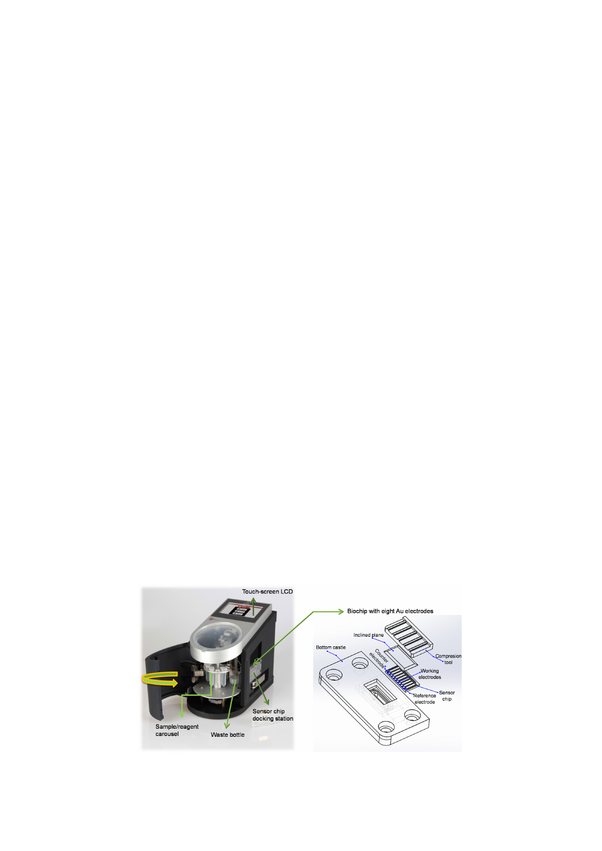
we designed and integrated a new chip (Figure 1) to improve the efficiency of the biosensing device and
the reproducibility of the assays. The electrodes were designed on the glass slide (10
×
20 mm) using a fine
metal mask, which was made of a laser-cut patterned stainless steel. Au was deposited on the wafer by
means of an electron beam evaporator (Ebeam system Nanovak NVEB-600, Nanovak, Ankara, Turkey).
Prior to the application of Au (200 nm), a 40 nm Ti layer was applied on to the wafer as an intermediary
adhesive layer to enhance the adhesion between the glass slide and the Au. Each array contains eight
working electrodes with a shared Au counter and quasi-reference electrodes. The experiments were
carried out using the MiCont
TM
software (TUB˙
ITAK-B˙
ILGEM, Kocaeli, Turkey) running on a wireless PC.
The assay protocols were generated, saved, and used when required.
Materials 2018, 11, x FOR PEER REVIEW 3 of 17
control surface probe: Biotin-5′- CAATATTTGGCGTGAATGGGTCGGAAAACA) for DNA sensor
development were obtained from Sentromer DNA Technologies LLC (Istanbul, Turkey).
2.2. Isolation of Salmonella Typhimurium from Human Stool Samples
Human stool sample was inoculated onto Salmonella-Shigella and xylose lysine deoxycholate
agar mediums, and incubated at 37 °C overnight. Bacterial culture was identified by using standard
biochemical tests (the use of glucose, citrate, and indole by bacteria, the production of gas and H
2
S
upon lactose fermentation of bacteria, the determination of the mobility and the ability to split urea).
The cultured colonies used glucose, citrate and indole confirmed the presence of Salmonella bacteria.
The isolate was serogrouped and serotyped using polyvalent and monovalent Salmonella antisera
(Statens Serum Institut, Copenhagen, Denmark) according to the Kauffmann-White scheme [26]. The
colonies that produced H
2
S were considered as a pure colony based on their morphology. After the
confirmation and serotyping, the strain was immediately frozen in tryptic soy broth with 16%
glycerol at −80 °C.
2.3. Fully-Automated Microfluidic-Based Electrochemical Sensor with a New Chip Design
A custom-designed fully-automated electrochemical sensor was used in this study to develop
antibody- and DNA-based biosensors for Salmonella detection. The sensor fabrication and its
developmental stages were fully described in our earlier studies [18,27,28]. Meanwhile, we have
realized several drawbacks dealing with device’s electronics and mechanics. One of the main
problems was electromagnetic noise in the signal. In order to decrease the noise level, two
modifications were employed: (1) Increasing of the distance between the microfluidic pumps and the
potentiometer. The microfluidic pump spread unwanted magnetic pulses by the pulse width
modulation (PWM) mechanism. (2) Applying a coaxial cable structure instead of unscreened cable
structure between the biosensor and potentiostat circuit. Herein, we designed and integrated a new
chip (Figure 1) to improve the efficiency of the biosensing device and the reproducibility of the assays.
The electrodes were designed on the glass slide (10 × 20 mm) using a fine metal mask, which was
made of a laser-cut patterned stainless steel. Au was deposited on the wafer by means of an electron
beam evaporator (Ebeam system Nanovak NVEB-600, Nanovak, Ankara, Turkey). Prior to the
application of Au (200 nm), a 40 nm Ti layer was applied on to the wafer as an intermediary adhesive
layer to enhance the adhesion between the glass slide and the Au. Each array contains eight working
electrodes with a shared Au counter and quasi-reference electrodes. The experiments were carried
out using the MiCont
TM
software (TUBİTAK-BİLGEM, Kocaeli, Turkey) running on a wireless PC.
The assay protocols were generated, saved, and used when required.
Figure 1. Illustration of MiSens electrochemical sensor and its new chip design.
Figure 1. Illustration of MiSens electrochemical sensor and its new chip design.
Materials 2018,11, 1541 4 of 17
2.4. Sensor Chip Cleaning and SAM Deposition
Prior to forming a self-assembled monolayer (SAM) on the sensor chip, the electrode surfaces were
cleaned by employing nitrogen plasma [
29
,
30
]. A 2 mM concentration of MUDA was used to prepare
the thiol solution in absolute ethanol for SAM deposition [
18
]. The sensor chips were immersed in the
ethanolic solution for overnight followed by washing with ethanol and Milli-Q water, respectively.
Later on the sensor chips were dried thoroughly under a stream of nitrogen gas, vacuum-packed,
and stored at +4 ◦C till use.
2.5. Selection of HRP Concentration for Bioassays
Different concentrations of HRP were initially measured using MiSens device (TUB˙
ITAK-B˙
ILGEM,
Kocaeli, Turkey) to determine the optimum HRP amount for bioassays. For this, six different
concentrations of HRP were mixed with same amount of TMB and injected to the MUDA coated
surfaces. These optimization experiments were repeated three times and the optimum HRP
concentration was chosen based on the average sensor signals.
2.6. Characterization of SAM Coated Sensor Chips Using AFM
The bare gold, the SAM coated and the antibody immobilized sensor chips were visualized
by employing a Naio (Nanosurf AG, Liestal, Switzerland) atomic force microscope (AFM).
Commercially available AFM probes (NCLR) from NanoWorld (NanoWorld AG, Liestal, Switzerland)
were used for AFM measurements. The AFM analyses for the SAM-deposited surface and the antibody
immobilization were carried out after having completed three cycles of buffer wash flow. The sensor
surfaces during the DNA assays were also visualized using AFM. Hence, the NA immobilization,
the capturing of DNA surface probe on the NA layer, and the target DNA binding were confirmed.
The chips were undocked from the MiSens sensor after three cycles of buffer flow, dried using a gentle
nitrogen stream, and then immediately placed in a glass Petri dish. All AFM measurements were
performed at room temperature using the intermittent air mode.
2.7. Development of the Antibody Biosensor
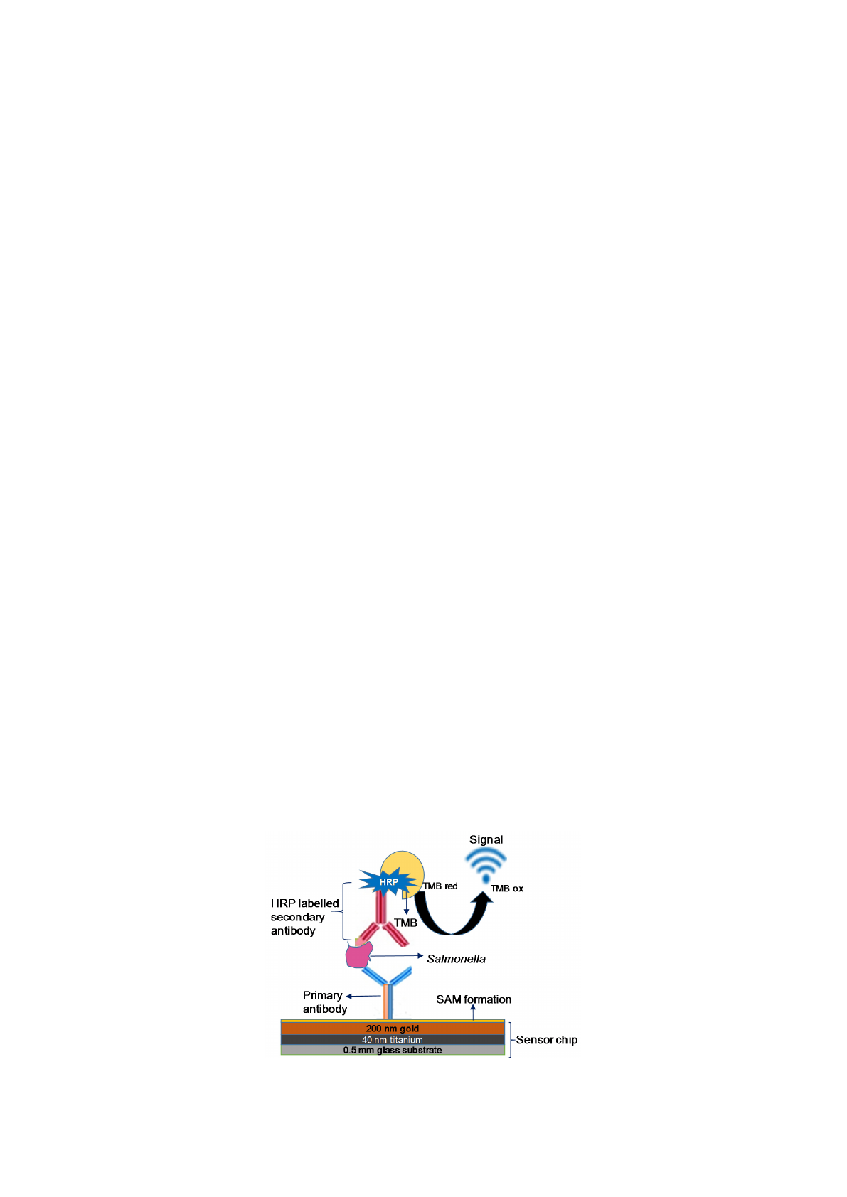
Two different antibody sensors were developed in this work by using a monoclonal primary
antibody and a secondary polyclonal antibody for Salmonella. The primary antibody was used as
a surface ligand to specifically capture Salmonella bacterium, whereas the polyclonal antibody was
utilized as a detector antibody to increase the sensor signal. Two different bio-detection assays
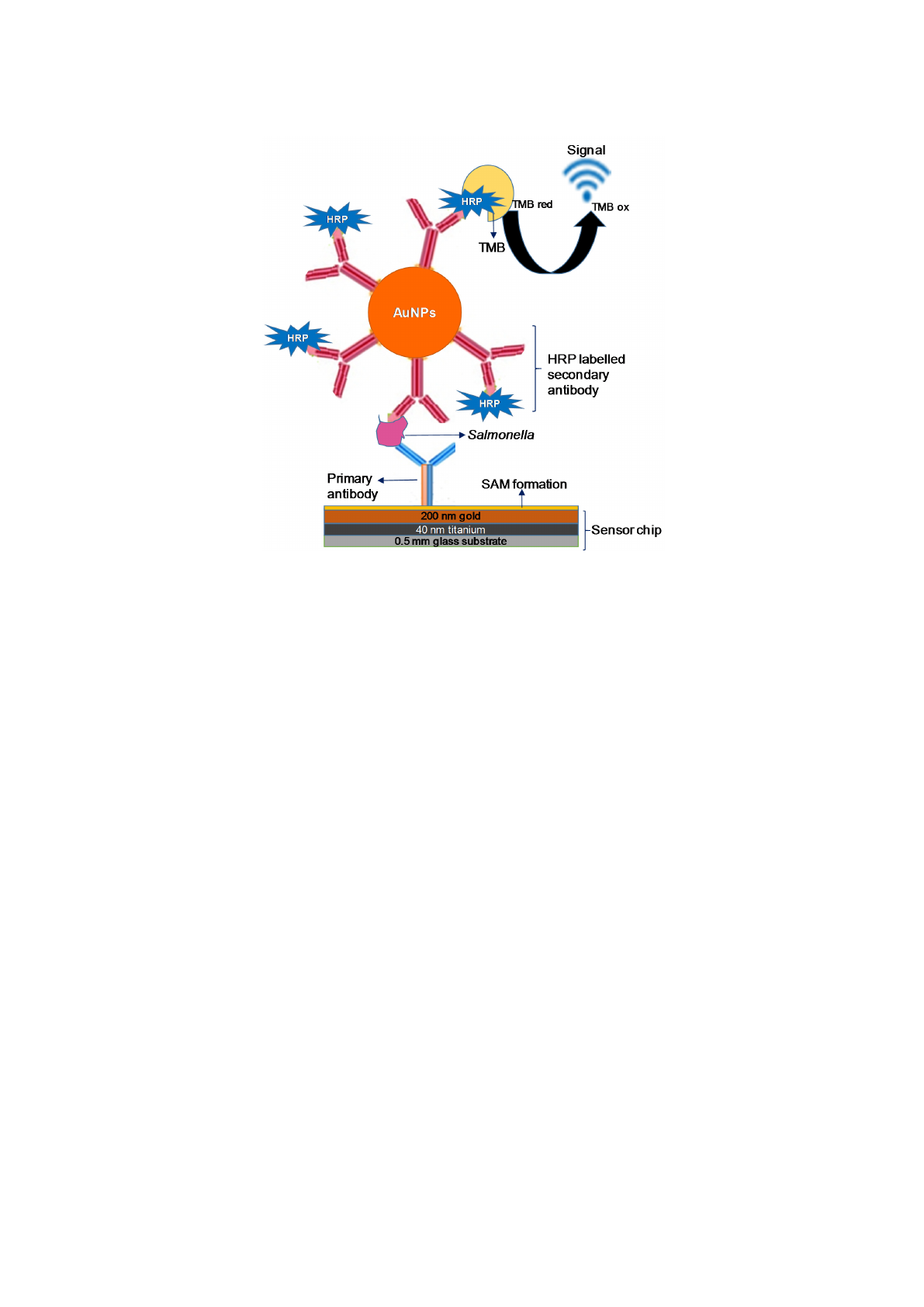
were established: a standard sandwich assay (Scheme 1) and a nanoparticle enhanced sensor assay
(Scheme 2). In the latter case the polyclonal antibodies were initially conjugated with gold nanoparticles
prior to the Salmonella detection assay.
Materials 2018, 11, x FOR PEER REVIEW 4 of 17
2.4. Sensor Chip Cleaning and SAM Deposition
Prior to forming a self-assembled monolayer (SAM) on the sensor chip, the electrode surfaces
were cleaned by employing nitrogen plasma [29,30]. A 2 mM concentration of MUDA was used to
prepare the thiol solution in absolute ethanol for SAM deposition [18]. The sensor chips were
immersed in the ethanolic solution for overnight followed by washing with ethanol and Milli-Q
water, respectively. Later on the sensor chips were dried thoroughly under a stream of nitrogen gas,
vacuum-packed, and stored at +4 °C till use.
2.5. Selection of HRP Concentration for Bioassays
Different concentrations of HRP were initially measured using MiSens device (TUBİTAK-
BİLGEM, Kocaeli, Turkey) to determine the optimum HRP amount for bioassays. For this, six
different concentrations of HRP were mixed with same amount of TMB and injected to the MUDA
coated surfaces. These optimization experiments were repeated three times and the optimum HRP
concentration was chosen based on the average sensor signals.
2.6. Characterization of SAM Coated Sensor Chips Using AFM
The bare gold, the SAM coated and the antibody immobilized sensor chips were visualized by
employing a Naio (Nanosurf AG, Liestal, Switzerland) atomic force microscope (AFM).
Commercially available AFM probes (NCLR) from NanoWorld (NanoWorld AG, Liestal,
Switzerland) were used for AFM measurements. The AFM analyses for the SAM-deposited surface
and the antibody immobilization were carried out after having completed three cycles of buffer wash
flow. The sensor surfaces during the DNA assays were also visualized using AFM. Hence, the NA
immobilization, the capturing of DNA surface probe on the NA layer, and the target DNA binding
were confirmed. The chips were undocked from the MiSens sensor after three cycles of buffer flow,
dried using a gentle nitrogen stream, and then immediately placed in a glass Petri dish. All AFM
measurements were performed at room temperature using the intermittent air mode.
2.7. Development of the Antibody Biosensor
Two different antibody sensors were developed in this work by using a monoclonal primary
antibody and a secondary polyclonal antibody for Salmonella. The primary antibody was used as a
surface ligand to specifically capture Salmonella bacterium, whereas the polyclonal antibody was
utilized as a detector antibody to increase the sensor signal. Two different bio-detection assays were
established: a standard sandwich assay (Scheme 1) and a nanoparticle enhanced sensor assay
(Scheme 2). In the latter case the polyclonal antibodies were initially conjugated with gold
nanoparticles prior to the Salmonella detection assay.
Scheme 1. Principle of antibody sensor development for Salmonella detection using the standard assay.
Scheme 1.
Principle of antibody sensor development for Salmonella detection using the standard assay.
Materials 2018,11, 1541 5 of 17
Materials 2018, 11, x FOR PEER REVIEW 5 of 17
The running buffer used for immobilization was degassed buffered saline (PBS, pH 7.4) and this
buffer continuously flowed over the Au sensor surfaces between the injections. The flow rate of the
buffer/reagents for the assay was 50 µL min
−1
unless written otherwise. For the assays, the SAM
deposited sensor chip was initially inserted to the device and primed with the running buffer (PBS).
The sensor surface was activated with a mixture of EDC (0.4 M) and NHS (0.1 M) in a 1:1 volume
ratio by 4 min injection prior to the covalent immobilization of the primary antibody (50 µg mL
−1
,
prepared in NaAc buffer, pH: 4.5) during the 4 min injection (50 µL min
−1
, 200 µL). The antibody-free
areas of the sensing surface were then blocked using a 100 µg mL
−1
BSA solution (50 µL min
−1
, 200
µL) and 1 M of ethanolamine (pH: 8.5, 50 µL min
−1
, 200 µL), respectively. Bio-detection capacity of
the sensor was tested in the investigation range of 1‒5.41 × 10
7
cfu mL
−1
using two different Salmonella
sample sources: commercially available S. typhimurium and real S. typhimurium samples from human
stool. The samples were prepared in a particular PBS buffer containing 200 µg mL
−1
BSA, 0.5 M NaCl,
500 µg mL
−1
dextran, and 0.5% Tween 20. Each sample was injected to the sensor surface (50 µL min
−1
,
200 µL) followed by the injection of the HRP-labelled secondary antibody (50 µL min
−1
, 200 µL).
Amperometric measurements were then carried out at −0.1 V using TMB reagent (4 min, 50 µL min
−1
)
followed by buffer injection (4 min, 100 µL min
−1
). The sensor surface regeneration was achieved with
the injection of 0.1 M HCl (1 min, 100 µL min
−1
) twice for the sequential detection of different
Salmonella concentrations.
The gold nanoparticle-enhanced bioassays (Scheme 2) were performed using the same
procedure. In this case, the HRP-labelled secondary antibody was conjugated with AuNPs prior to
the experiments to amplify the sensor signal. The AuNP conjugation protocol was described in our
earlier studies [18] and used as is in this work. The secondary antibody-labelled AuNPs were stored
at +4 °C and warmed to room temperature before use. The concentration of nanoparticles was
calculated at 525 nm wavelength and the AuNP solution was diluted based on the dilution factor
calculated by considering the OD value. Commercially available S. typhimurium and real S.
typhimurium samples from human stool were detected in a concentration range from 1 to 5.41 × 10
7
cfu mL
−1
using the amperometric sensor.
Scheme 2. Principle of the gold nanoparticles enhanced immunosensor for Salmonella detection.
Scheme 2. Principle of the gold nanoparticles enhanced immunosensor for Salmonella detection.
The running buffer used for immobilization was degassed buffered saline (PBS, pH 7.4) and this
buffer continuously flowed over the Au sensor surfaces between the injections. The flow rate of the
buffer/reagents for the assay was 50
µ
L min
−1
unless written otherwise. For the assays, the SAM
deposited sensor chip was initially inserted to the device and primed with the running buffer (PBS).
The sensor surface was activated with a mixture of EDC (0.4 M) and NHS (0.1 M) in a 1:1 volume
ratio by 4 min injection prior to the covalent immobilization of the primary antibody (50
µ
g mL
−1
,
prepared in NaAc buffer, pH: 4.5) during the 4 min injection (50
µ
L min
−1
, 200
µ
L). The antibody-free
areas of the sensing surface were then blocked using a 100
µ
g mL
−1
BSA solution (50
µ
L min
−1
, 200
µ
L)
and 1 M of ethanolamine (pH: 8.5, 50
µ
L min
−1
, 200
µ
L), respectively. Bio-detection capacity of the
sensor was tested in the investigation range of 1–5.41
×
10
7
cfu mL
−1
using two different Salmonella
sample sources: commercially available S. typhimurium and real S. typhimurium samples from human
stool. The samples were prepared in a particular PBS buffer containing 200
µ
g mL
−1
BSA, 0.5 M
NaCl, 500
µ
g mL
−1
dextran, and 0.5% Tween 20. Each sample was injected to the sensor surface
(50
µ
L min
−1
, 200
µ
L) followed by the injection of the HRP-labelled secondary antibody (50
µ
L min
−1
,
200
µ
L). Amperometric measurements were then carried out at
−
0.1 V using TMB reagent (4 min,
50
µ
L min
−1
) followed by buffer injection (4 min, 100
µ
L min
−1
). The sensor surface regeneration was
achieved with the injection of 0.1 M HCl (1 min, 100
µ
L min
−1
) twice for the sequential detection of
different Salmonella concentrations.
The gold nanoparticle-enhanced bioassays (Scheme 2) were performed using the same procedure.
In this case, the HRP-labelled secondary antibody was conjugated with AuNPs prior to the experiments
to amplify the sensor signal. The AuNP conjugation protocol was described in our earlier studies [
18
]
and used as is in this work. The secondary antibody-labelled AuNPs were stored at +4
◦
C and
warmed to room temperature before use. The concentration of nanoparticles was calculated at
525 nm wavelength and the AuNP solution was diluted based on the dilution factor calculated by
considering the OD value. Commercially available S. typhimurium and real S. typhimurium samples
from human stool were detected in a concentration range from 1 to 5.41
×
10
7
cfu mL
−1
using the
amperometric sensor.
Loading more pages...