
Toxicology Reports 7 (2020) 1578–1587
Available online 21 November 2020
2214-7500/© 2020 The Authors. Published by Elsevier B.V. This is an open access article under the CC BY license (http://creativecommons.org/licenses/by/4.0/).
3D-bioprinted HepaRG cultures as a model for testing long term aflatoxin
B1 toxicity in vitro
Konrad Schmidt, Johanna Berg, Viola Roehrs, Jens Kurreck, Munir A. Al-Zeer*
Department of Applied Biochemistry, Institute of Biotechnology, 4/3-2, Technische Universit¨
at Berlin, Gustav-Meyer-Allee 25, 13355 Berlin, Germany
ARTICLE INFO
Keywords:
3D-bioprinting
Aflatoxin B1
Alternative in vitro model
HepaRG liver cells
ABSTRACT
In recent years 3D-bioprinting technology has been developed as an alternative to animal testing. It possesses a
great potential for in vitro testing as it aims to mimic human organs and physiology. In the present study, an
alginate-gelatin-Matrigel based hydrogel was used to prepare 3D-bioprinted HepaRG cultures using a pneumatic
extrusion printer. These 3D models were tested for viability and metabolic functions. Using 3D-bioprinted
HepaRG cultures, we tested the toxicity of aflatoxin B1 (10 or 20
μ
M) in vitro and compared the results with
2D HepaRG cultures. There was a dose-dependent toxicity effect on cell viability, reduction of metabolic activity
and albumin production. We found that 3D-bioprinted HepaRG cultures are more resistant to aflatoxin B1
treatment than 2D cultures. Although the metabolic activities were reduced upon treatment with aflatoxin B1,
the 3D models were still viable and survived longer, up to 3 weeks, than the 2D culture, as visualized by fluo-
rescence microscopy. Furthermore, albumin production recovered slightly in 3D models after one and two weeks
of treatment. Taken together, we consider using 3D-bioprinting technology to generate 3D tissue models as an
alternative way to study toxicity in vitro and this could also provide a suitable alternative for chronic hepato-
toxicity studies in vitro.
1. Introduction
In vitro cell cultures and animal experiment models are crucial in-
struments in basic research and preclinical studies [1–3]. Even though
cell cultures and animal models are widely used in the field of toxicology
testing, recent advances in 3D-bioprinting technology are emerging as a
useful tool for testing of complex cell environments, toxicity and for
drug discovery in vitro [4,5,42] . Traditional 2D cell culture systems
using cell lines or primary cells do not recapitulate the natural
three-dimensional microenvironment of the respective human tissue or
organ, which in turn leads to changes in gene and protein expression [1,
6]. On the other hand, beyond the inherent complexity of animal
models, the microenvironments of animal tissues do not efficiently
recapitulate many of their human counterparts [7]. The creation of
microenvironments in vitro that capture some features of human tissues
could be enabled by advances in the understanding of 3D-bioprinting
[8]. For this reason, 3D-bioprinting is considered a promising tool to
study infection, cancer, drug screening, and toxicity testing [9,10]. This
can reduce or replace the need for animal testing that does not precisely
reflect human responses in terms of toxicology and pathophysiology.
Although 3D-bioprinting is a cutting edge technology used to fabri-
cate 3D models in vitro that hold tremendous promise, especially in the
fields of biology and medicine, 3D-bioprinting technology and bio-
materials are still being fine-tuned [11]. Bioprinting offers great preci-
sion that enables the exact spatial and temporal deposition of biological
materials, including cells and extracellular matrix in a 3D hierarchal
organization to generate artificial 3D models of native tissues. The
precise deposition and spatial arrangement of these components can
recapitulate the natural architecture and the microenvironments of the
tissue in vitro [11]. Both natural and synthetic polymers, such as collagen
[12], gelatin [13], and alginate [14] etc., are used extensively to fabri-
cate scaffolds for 3D model printing due to on their biocompatibility,
printability and biodegradability properties [15].
Several 3D mini-organs and tissue constructs have been printed and
fabricated in vitro, including skin, heart, bone, cartilage, lung, neurons,
and pancreas using different scaffolds and utilizing various 3D-bio-
printing approaches [11]. Liver 3D mini-models have also been gener-
ated in vitro using various tissue engineering techniques including
3D-bioprinting [16]. Various cell models have been used to fabricate
3D liver model in vitro including the use of human iPSC-derived
* Corresponding author.
E-mail address: [email protected] (M.A. Al-Zeer).
Contents lists available at ScienceDirect
Toxicology Reports
journal homepage: www.elsevier.com/locate/toxrep
https://doi.org/10.1016/j.toxrep.2020.11.003
Received 14 July 2020; Received in revised form 4 November 2020; Accepted 5 November 2020

Toxicology Reports 7 (2020) 1578–1587
1579
hepatocytes [4], HepG2 cells [17], and HepaRG cell line [18]. The
HepaRG cell line has been shown to be stable in vitro, allowing long-term
culture [18]. Liver models using 3D-bioprinted cells are becoming more
common in clinical and basic research, including infection, drug dis-
covery, and toxicity testing [19,20]. As the liver plays a central role in
metabolism and detoxification of chemicals, 3D-bioprinting possesses
great potential to achieve these goals and model complexity that mimics
the liver functions in vitro [43]. Therefore, 3D-bioprining has the po-
tential to become an important tool for in vitro toxicity testing especially
for studies of chronic hepatotoxicity against toxins such as aflatoxins.
Aflatoxins are secondary fungal metabolites, known as mycotoxins,
which are mainly produced by the genus Aspergillus [21,22]. They are
classified as Group 1 carcinogen by the International Agency for
Research in Cancer [23]. Aflatoxins are hepatotoxic and have been
implicated in increasing the risk of hepatocellular carcinoma. Aflatoxin
B1 has been shown to induce cytotoxicity in HepaRG cells [44]. This
hepatotoxicity is mediated by its toxic epoxide, which is produced by
CYP1A2 and CYP3A4 [24]. Further, aflatoxin B1 induces loss of cell
viability due to the induction of apoptosis in HepaRG cells. This
phenotype has been correlated with DNA damage response mediated by
p53 signaling [24,25].
In this study, we aimed at optimizing a 3D liver cell model using
bioprinting technology with an alginate/gelatin/Matrigel-based scaf-
fold, which can be used for extrusion-based bioprinting of HepaRG cell
models for toxicity testing. The 3D liver constructs were generated using
a 3D micro-extrusion bioprinter. Viability, cytotoxicity and metabolic
activity of the printed constructs were evaluated. Upon treatment with
AFB1, cells grown in 2D did not survive the damage induced by the
toxin, unlike the 3D-bioprinted constructs. Although AFB1 reduced
metabolic activity in the 3D-bioprinted constructs, cells survived the
AFB1 toxicity and were still viable, as visualized by fluorescence mi-
croscopy. Therefore, a 3D-bioprinted model may pave the way to study
the long-term effect of AFB1 and carcinogenesis in vitro.
2. Materials and methods
2.1. Cell culture
HepaRG cells were obtained from Biopredic International (Saint
Gregoir´
e, France). The cells were cultured in William’s E medium
(Gibco, Dreieich, Germany) supplemented with 2 mM L-Glutamine
(Biowest, Nuaill´
e, France), 10 % fetal bovine serum (FBS; c.c.pro,
Oberdorla, Germany), 50
μ
M hydrocortisone hemisuccinate (Sigma,
Steinheim, Germany), 5
μ
g/mL recombinant human insulin (PAN
Biotech, Aidenbach, Germany), and 1 % penicillin/streptomycin (P/S;
Biowest). The cells were cultured at 37 ◦C and 5 % CO
2
in a humidified
incubator for 14 days before differentiation and the medium was
renewed every three days. After 14 days, hepatic differentiation was
induced by adding 1.7 % DMSO (Sigma) to the culture medium for
additional 14 days as previously described [20].
2.2. Preparation of cell-scaffold hydrogels
Sodium alginate (4.5 % w/v) and gelatin (6.5 % w/v) powders
(Sigma) were dissolved in William’s E medium on a magnetic stirrer at
1250 min
−1
(overnight at 37 ◦C). The hybrid alginate -gelatin hydrogel
(450
μ
l) was mixed with liquid Matrigel (200
μ
l) (Corning, Tewksbury,
MA, USA), differentiated HepaRG cells, 1.22 M CaSO
4
(25
μ
l) (Roth,
Karlsruhe, Germany) and William’s E medium containing supplements
(325
μ
l) to obtain the final cell-scaffold mixture bioink composed of
alginate (2 % w/v), gelatin (3 % w/v), Matrigel (20 % w/v), 30 mM
CaSO
4
and 7 * 10
6
HepaRG cells/ml, as previously described [26].
Following the initial cross-linking of alginate using CaSO
4
(8 min after
mixing), the cell-scaffold hydrogel was loaded into the printing cartridge
(Cellink, Gothenburg, Sweden).
2.3. 3D bioprinting
The Cellink INKREDIBLE+3D-printer was utilized for the bio-
printing process which was carried out at room temperature. A
rectangular-shaped construct (1 mm height x10 mm width x10 mm
length) was designed with regularly spaced pores in a grid-like pattern
using Slic3r software. The shape was selected to allow the diffusion of
nutrients, oxygen, and metabolites through the constructs. The hydrogel
was extruded through a 22 G needle at 10–40 kPa to generate 3D-liver
constructs designed by the computer-aided design (CAD) software Rhi-
noceros5 (Robert McNeel & Associates, Seattle, WA, USA). The printed
constructs were further cross-linked using 100 mM CaCl
2
(Roth) to in-
crease the gelation of alginate. Then, constructs were cultured with
William’s E medium supplemented with 1.7 % DMSO, as well as 20 mM
CaCl
2
, and incubated at 37 ◦C and 5 % CO
2
in a humidified incubator.
2.4. Staining with fluorescent DNA dye and immunofluorescence
Printed 3D constructs were fixed for 30 min using 4 % formaldehyde.
The constructs were then permeabilized with 1 % Triton-X-100 (Roth)
for 1 h at room temperature and the nuclei were stained with Hoechst
dye (1
μ
g/ml) (H33342, AppliChem, Darmstadt, Germany). Cellular
distribution was analyzed with the Zeiss Observer. Z1 microscope (Zeiss,
Jena, Germany).
2.5. The live/dead viability assay
To determine cell viability, we used a commercial Live/Dead assay
(Viability/Cytotoxicity kit; ThermoFisher Scientific, Waltham, MA,
USA). Briefly, the 3D liver constructs were incubated with 2
μ
M calcein-
AM and 4 mM ethidium homodimer-1 diluted in 1x HBSS (ThermoFisher
Scientific) for 15 min (37 ◦C, 5 % CO
2
). Subsequently, samples were
analyzed by fluorescence microscopy (Zeiss Observer. Z1 microscope;
Zeiss, Germany).
2.6. XTT assay
Metabolic activity of HepaRG cells printed constructs or 2D cultures
was determined using the tetrazolium hydroxide salt (XTT) assay ac-
cording to the manufacturer’s instructions (AppliChem, Germany) at
indicated time points. The absorbance at 450 nm, with a reference of
620 nm, was carried out using TriStar Multimode Reader LB942 (Bert-
hold Technologies, Bad Wildbad, Germany). Cell-laden constructs
incubated in 10 % Triton-X-100 (Roth), which was diluted culture me-
dium were used as lysis control. Values were normalized to the lysis
controls.
2.7. Lactate dehydrogenase release
The supernatants from different constructs were collected at each
time point, as indicated, and lactate dehydrogenase colorimetric assays
(Roche, Switzerland) were carried out according to the manufacturer’s
instructions. The absorbance at 490 nm, with a reference of 620 nm, was
measured with the Sunrise microplate reader (Tecan, M¨
annedorf,
Switzerland).
2.8. Albumin measurement
The supernatants from 3D constructs or 2D monolayers were
analyzed and quantified for albumin secretion using the human albumin
enzyme-linked immunosorbent assay (ELISA) kit (Bethyl Laboratories,
Montgomery, TX, USA), according to the manufacturer’s instructions.
The absorbance at 450 nm, with a reference of 620 nm, was carried out
using TriStar Multimode Reader LB942.
K. Schmidt et al.
Toxicology Reports 7 (2020) 1578–1587
1580
2.9. Treatment with toxins
Differentiated HepaRG cells were treated with 10, or 20
μ
M aflatoxin
B1 (AFB1) (Sigma-Aldrich). The AFB1 was dissolved in dimethylsulf-
oxide (DMSO). Control cells were treated with the same volume of
DMSO for 1, 2, 5 and 7 days of incubation. For long-term culture cells
were treated with single dose 10, or 20
μ
M AFB1 for one week then cells
were cultured for additional two weeks without the toxin. A single dose
of doxorubicin (Dox) (Sigma-Aldrich) was used to treat the constructs
for 1, 2, and 3 days. Dox was also dissolved in DMSO to a final con-
centration of 10, or 20
μ
M.
2.10. Statistical analysis
Data were analyzed using Student’s t-test (GraphPad Prism 6,
GraphPad Software, Inc., La Jolla, CA, USA). Data are represented as
mean ±SD, p-values are considered significant by *p ≤0.05; **p ≤0.01;
***p ≤0.001; **** p ≤0.0001.
3. Results
3.1. Preparation and characterization of HepaRG cell-scaffold bioink
Bioink optimization is a critical step in bioprinting. To assess the
suitability of the hydrogel composed of alginate-gelatin-Matrigel (2 %, 3
%, and 20 % respectively) for 3D-bioprinting, differentiated HepaRG
cells were used to generate 3D constructs for toxicity testing studies in
vitro. The preparation of the printing process was performed as previ-
ously described [26]. Briefly, the hydrogel was pre-heated to 37 ◦C and
loaded into a three ml-syringe, cells, Matrigel and CaSO
4
were loaded
Fig. 1. Characterization of bioprinted 3D constructs. (A) Qualitative viability staining of the 3D printed mature HepaRG cells in the constructs at the indicating time
points using the Live/Dead staining; calcein-AM (live cells in green) and ethidium homodimer-1 (dead cells in red). (B) Spatial distribution of mature HepaRG cells in
3D constructs using alginate/gelatin/Matrigel as a bioink at the following time points: 1, 7, 14, and 21 days post-printing visualized by nuclear Hoechst staining
(blue). Graphs are representative images of three independent experiments. (C) Metabolic activity of mature HepaRG cells in the 3D constructs was determined by
tetrazolium hydroxide salt (XTT) assays at the indicated time points post-printing. Absorbance was carried out at 450 nm. (D) Albumin secretion of mature HepaRG
3D constructs quantified using enzyme-linked immunosorbent assay (ELISA) analysis at the indicated time points. Results are shown as mean ±SD of three inde-
pendent experiments. * p ≤0.05; ** p ≤0.01; *** p ≤0.001; **** p ≤0.0001.
K. Schmidt et al.
Toxicology Reports 7 (2020) 1578–1587
1581
into another syringe. Using a Luer-lock adapter, both syringes were
connected, and the hydrogels mixed thoroughly. They were then loaded
onto a printing cartridge and printed using the INKREDIBLE +bio-
printer (Cellink). The optimized hydrogel consisting of
alginate-gelatin-Matrigel enabled the printing procedure, and protected
the integrity of the 3D constructs, cell viability, and metabolic activity of
the cells.
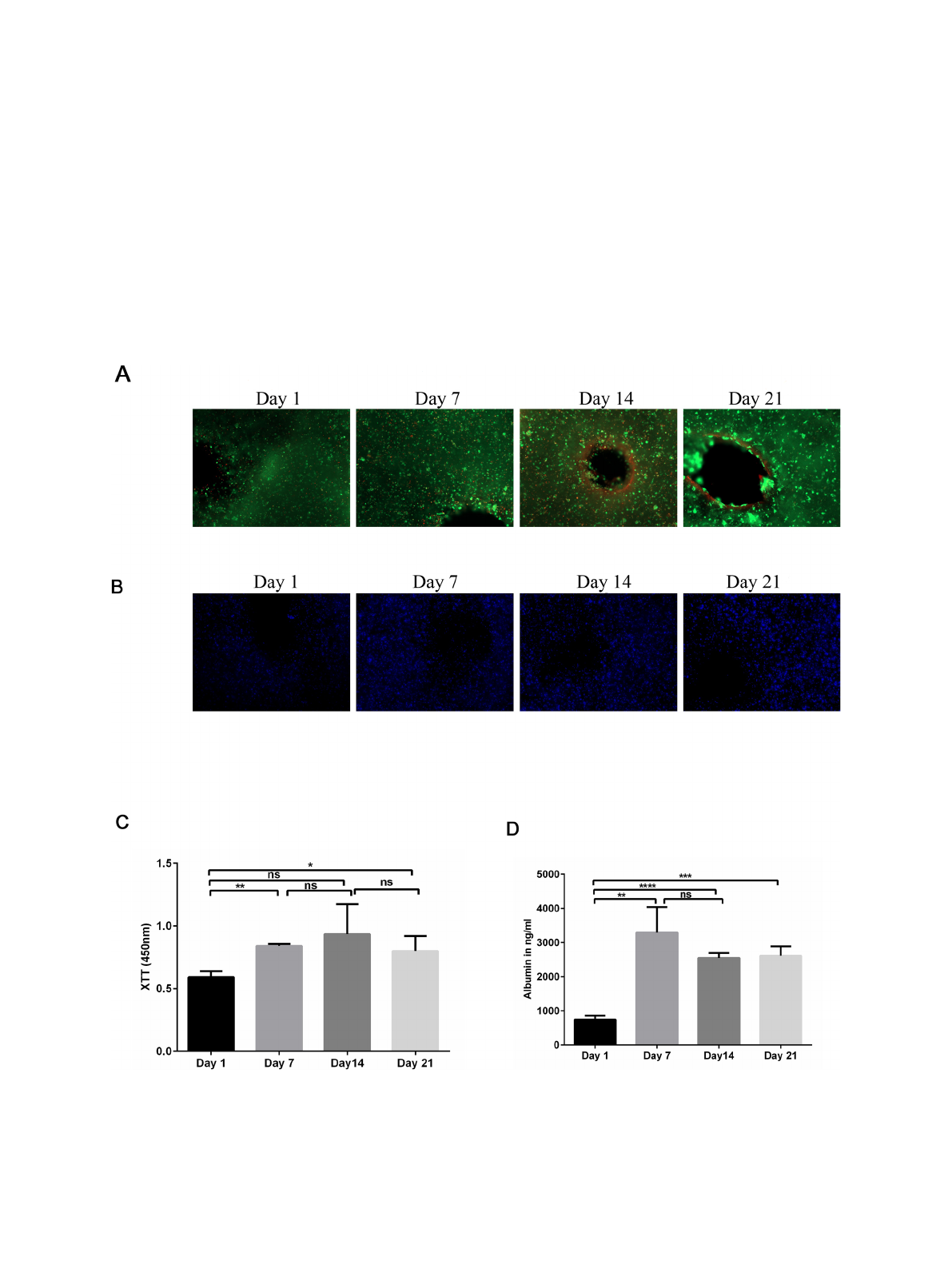
The viability of the printed cells was confirmed by live/dead staining
(Fig. 1A). The 3D constructs were qualitatively analyzed using calcein-
AM (green) to stain living cells and ethidium homodimer-1 (red) to
stain dead cells after 1, 7, 14, and 21 days of culture, followed by
fluorescence microscopy. No obvious increase in the number of dead,
ethidium homodimer-1-positive cells, was detected at any of the time
points. Furthermore, the number of living, calcein-AM-positive cells,
was relatively stable during the entire time course (Fig. 1A). Further, we
analyzed the spatial distribution of the differentiated HepaRG cells
within the 3D constructs using fluorescence microscopy. First, the nuclei
were stained with Hoechst DNA dye (blue) and the distribution of the
cells was visualized at the indicated time points (Fig. 1B). Cells were
homogeneously distributed in the printed 3D constructs without sig-
nificant differences during the time course of the experiment (Data not
shown). Taken together, we conclude that the 3D-printed liver con-
structs are viable, and the cells are distributed evenly throughout the
hydrogel.
Using XTT assays, we quantified the metabolic activity of the Hep-
aRG cells in the 3D printed constructs (Fig. 1C). Consistent with the cell
viability obtained using the live/dead assay, the metabolic activity in
the 3D constructs increased significantly at day 7 and 21 compared to
day 1 (Fig. 1C). Furthermore, we analyzed albumin secretion using
ELISAs to determine the metabolic activity of the hepatic cells in 3D
constructs (Fig. 1D). Albumin secretion is one of the main characteristics
of hepatocytes and its production reflects a specific-function of liver
cells. The amount of albumin secreted from 3D printed cells increased
over time, and this increase was statistically significant at days 7, 14 and
21 (Fig. 1D). Taken together, our results showed substantial increases in
cell viability and metabolic activity over time in the 3D cell culture
system.
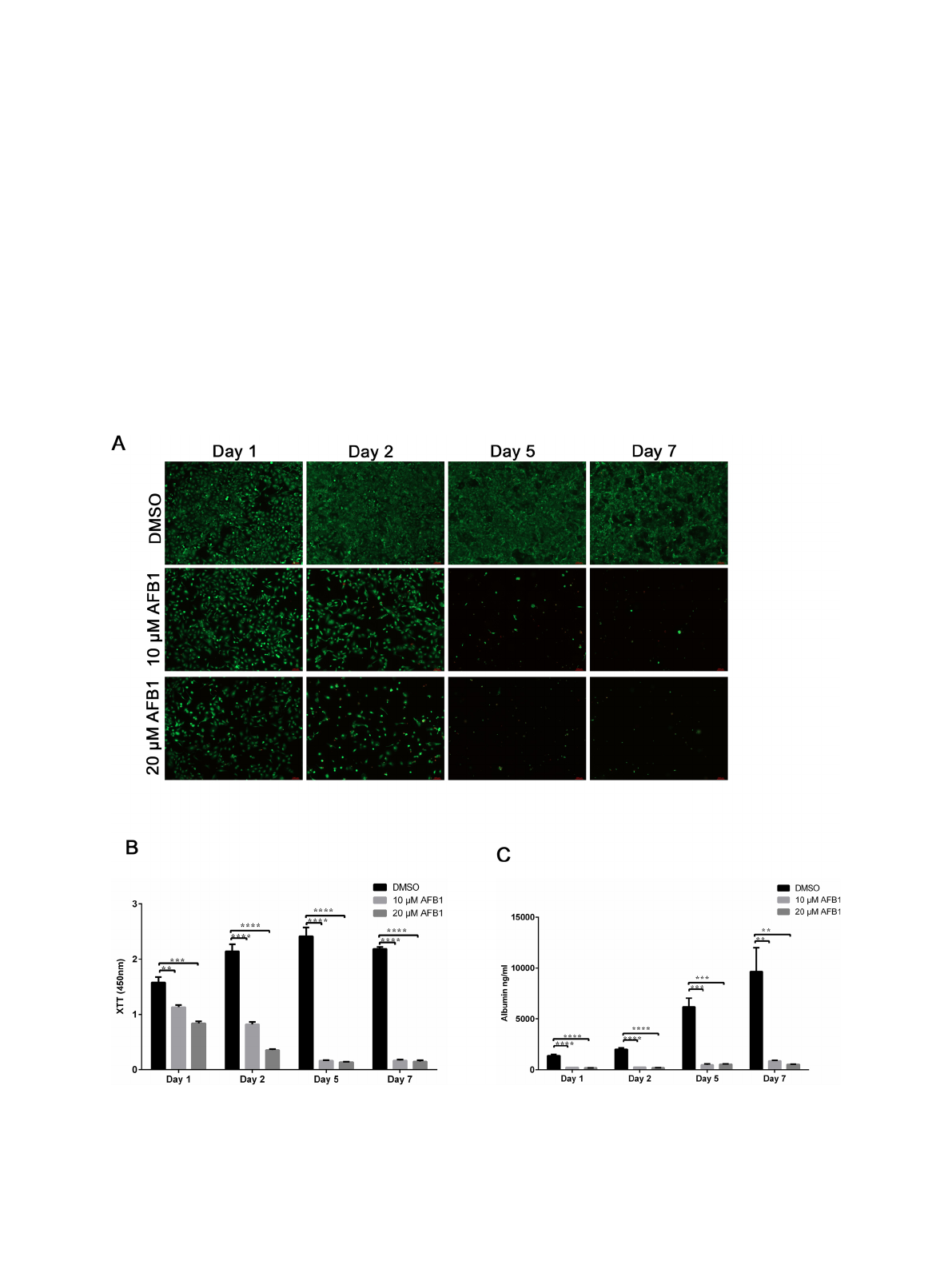
Fig. 2. Assessment of aflatoxin B1 toxicity on liver cells grown as 2D monolayers. (A) Monolayers of mature HepaRG cells, treated with DMSO, 10
μ
M AFB1, and 20
μ
M AFB1 at the indicated time points were tested for cell survival using live-dead staining. Live cells fluoresce green, whereas dead cells fluoresce red. Graphs are
representative images of three independent experiments. (B) Metabolic activity of mature HepaRG cells grown in 2D upon AFB1 treatment at the indicated time
points. Activities were determined using tetrazolium hydroxide salt (XTT) assays. Absorbance was carried out at 450 nm. (C) Albumin levels of mature HepaRG cells
in 2D monolayers upon AFB1 exposure were quantified at the indicated time points using ELISA analysis. Results are shown as mean ±SD of three independent
experiments. * p ≤0.05; ** p ≤0.01; *** p ≤0.001; **** p ≤0.0001.
K. Schmidt et al.
Toxicology Reports 7 (2020) 1578–1587
1582
3.2. Assessment of aflatoxin B1 toxicity using HepaRG 2D culture
To study the sensitivity of cells cultured in 2D to toxicity stemming
from AFB1, HepaRG cells were seeded onto a12-well plate at a density of
5*10
5
cells per well. Cells were then treated with a single dose of AFB1
(10 and 20
μ
M) or DMSO, which served as a control. Following treat-
ment with AFB1, cells were stained and analyzed for cell viability using
live/dead stain and fluorescence microscopy. AFB1 treatment (10
μ
M)
resulted in a reduction of viable cells (calcein-AM positive-cells shown in
green) after 24 and 48 h (Fig. 2A). Similar results were obtained from the
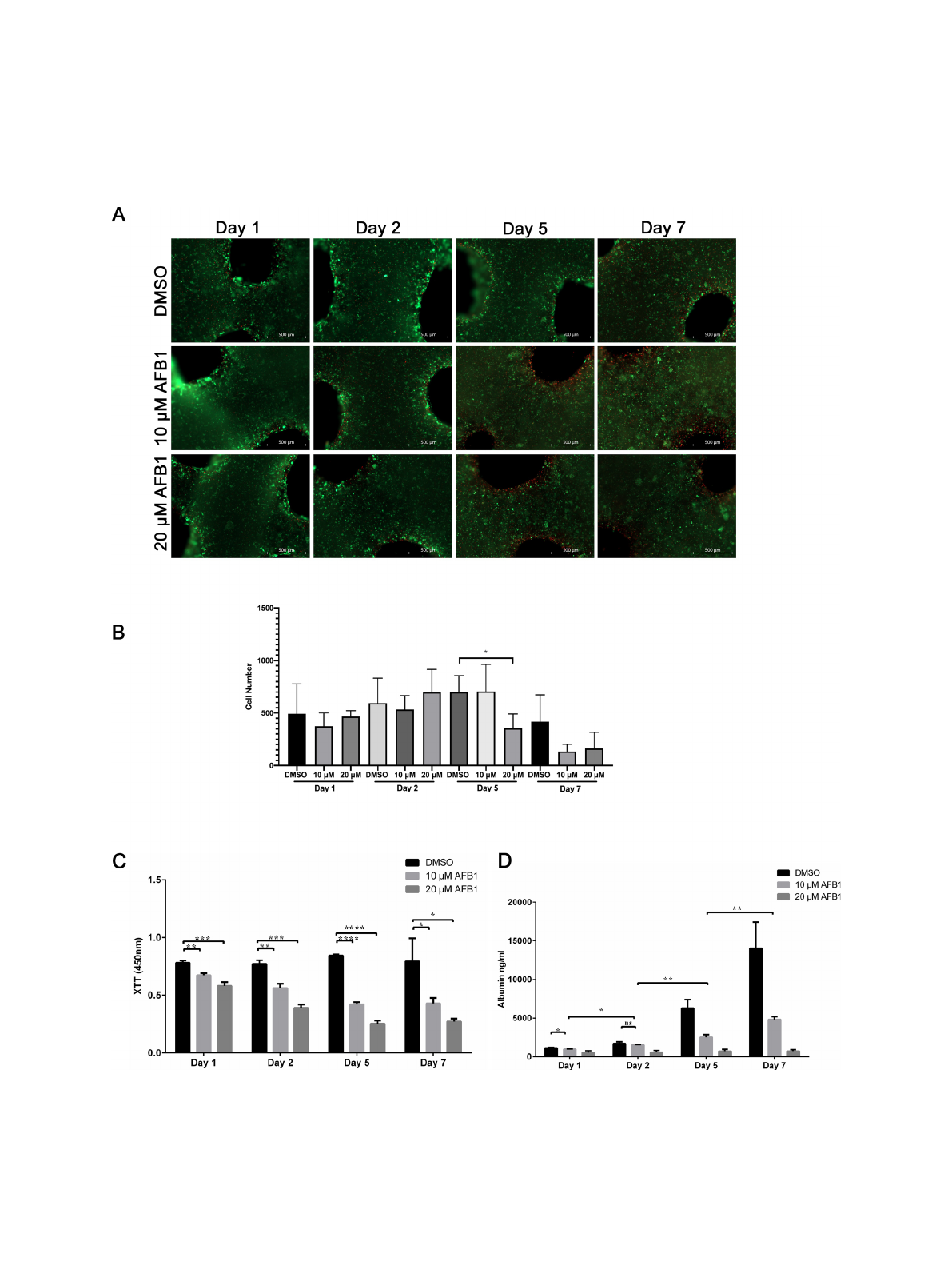
Fig. 3. Effect of aflatoxin B1 on liver cells in 3D printed constructs. (A) Cell viability of mature HepaRG in 3D constructs upon treatment with AFB1 (10
μ
M or 20
μ
M)
for the indicated time periods was visualized using the live-dead staining; calcein-AM (living cells in green) and ethidium homodimer-1 (dead cells in red). (B)
Number of green-stained cells quantified with ImageJ in constructs treated with AFB1 for the indicated time points. Bars indicate the means ±SD, n ≥3 images. (C)
Metabolic activity of the mature HepaRG cells treated with DMSO or AFB1 (10
μ
M or 20
μ
M) inside the 3D printed alginate/gelatin/Matrigel constructs was
determined by the XTT assay at the indicated time points. Absorbance was measured at 450 nm. (D) Levels of albumin secreted from mature HepaRG cells in 3D
constructs upon DMSO or AFB1 (10
μ
M or 20
μ
M) treatment were quantified at the indicated time points using ELISA analysis. Results are shown as mean ±SD of
three independent experiments. * p ≤0.05; ** p ≤0.01; *** p ≤0.001; **** p ≤0.0001.
K. Schmidt et al.
Loading more pages...