
Adrian Jonas, Steffen Staeck, Birgit Kanngießer, Holger Stiel, Ioanna
Mantouvalou
Laboratory quick near edge x-ray absorption fine
structure spectroscopy in the soft x-ray range with
100 Hz frame rate using CMOS technology
Open Access via institutional repository of Technische Universität Berlin
Document type
Journal article | Published version
(i. e. publisher-created published version, that has been (peer-) reviewed and copyedited; also known as:
Version of Record (VOR), Final Published Version)
This version is available at
https://doi.org/10.14279/depositonce-15602
Citation details
This article may be downloaded for personal use only. Any other use requires prior permission of the author
and AIP Publishing. This article appeared in
Jonas, A., Staeck, S., Kanngießer, B., Stiel, H., & Mantouvalou, I. (2021). Laboratory quick near edge x-ray
absorption fine structure spectroscopy in the soft x-ray range with 100 Hz frame rate using CMOS technology.
In Review of Scientific Instruments (Vol. 92, Issue 2, p. 023102). AIP Publishing.
and may be found at https://doi.org/10.1063/5.0032628.
Terms of use
This work is protected by copyright and/or related rights. You are free to use this work in any way permitted by
the copyright and related rights legislation that applies to your usage. For other uses, you must obtain
permission from the rights-holder(s).
Rev. Sci. Instrum. 92, 023102 (2021); https://doi.org/10.1063/5.0032628 92, 023102
© 2021 Author(s).
Laboratory quick near edge x-ray
absorption fine structure spectroscopy in
the soft x-ray range with 100Hz frame rate
using CMOS technology
Cite as: Rev. Sci. Instrum. 92, 023102 (2021); https://doi.org/10.1063/5.0032628
Submitted: 12 October 2020 • Accepted: 24 January 2021 • Published Online: 16 February 2021
Adrian Jonas, Steffen Staeck, Birgit Kanngießer, et al.
ARTICLES YOU MAY BE INTERESTED IN
Attosecond state-resolved carrier motion in quantum materials probed by soft x-ray
XANES
Applied Physics Reviews 8, 011408 (2021); https://doi.org/10.1063/5.0020649
High-resolution inelastic x-ray scattering at the high energy density scientific instrument at
the European X-Ray Free-Electron Laser
Review of Scientific Instruments 92, 013101 (2021); https://doi.org/10.1063/5.0022886
Determining the water content of nominally anhydrous minerals at the nanometre scale
Review of Scientific Instruments 92, 023103 (2021); https://doi.org/10.1063/5.0025570

Review of
Scientific Instruments ARTICLE scitation.org/journal/rsi
Laboratory quick near edge x-ray absorption fine
structure spectroscopy in the soft x-ray range
with 100Hz frame rate using CMOS technology
Cite as: Rev. Sci. Instrum. 92, 023102 (2021); doi: 10.1063/5.0032628
Submitted: 12 October 2020 •Accepted: 24 January 2021 •
Published Online: 16 February 2021
Adrian Jonas,1,2,a) Steffen Staeck,1,2 Birgit Kanngießer,1,2 Holger Stiel,1,3 and Ioanna Mantouvalou1,2,b)
AFFILIATIONS
1Berlin Laboratory for Innovative X-ray Technologies (BLiX), D-10623 Berlin, Germany
2TU Berlin, Analytical X-Ray Physics, D-10623 Berlin, Germany
3Max-Born-Institut für Nichtlineare Optik und Kurzzeitspektroskopie, D-12489 Berlin, Germany
a)Author to whom correspondence should be addressed: [email protected]
b)Current address: Helmholtz Zentrum Berlin, D-12489, Germany.
ABSTRACT
In laboratory based x-ray absorption fine structure (XAFS) spectroscopy, the slow readout speed of conventional CCD cameras can prolong
the measuring times by multiple orders of magnitude. Using pulsed sources, e.g., laser-based x-ray sources, the pulse repetition rate often
exceeds the frame rate of the CCD camera. We report the use of a scientific CMOS (sCMOS) camera for XAFS spectroscopy with a laser-
produced plasma source facilitating measurements at 100Hz. With this technological improvement, a new class of experiments becomes
possible, starting from the time consuming analysis of samples with small absorption to pump-probe investigations. Furthermore, laboratory
quicksoftx-rayabsorptionfinestructure(QXAFS)measurementswith10mstime resolution are rendered feasible. We present the character-
ization of the sCMOS camera concerning noise characteristics and a comparison to conventional CCD camera performance. The feasibility
of time resolved QXAFS measurements is shown by analyzing the statistical uncertainty of single shot spectra. Finally, XAFS spectroscopy on
a complex sandwich structure with minute amounts of NiO exemplifies the additional merits of fast detectors.
Published under license by AIP Publishing. https://doi.org/10.1063/5.0032628
I. INTRODUCTION
X-ray absorption fine structure (XAFS) spectroscopy is an
analytical tool for the investigation of the element specific elec-
tronic structure of atoms or molecules inside a sample. In quick
x-ray absorption fine structure (QXAFS) measurements, absorption
spectra are taken in quick succession to resolve dynamical pro-
cesses in the millisecond to minute range.1–10 The technique is a
powerful tool for investigating in situ reactions and slow dynamic
processes. XAFS is currently most commonly performed at syn-
chrotron radiation facilities due to the available high flux and effi-
cient instrumentation. For measurements, the excitation energy is
scanned in the region of the probed absorption edge and absorption
is measured via transmission, fluorescence, or electron detection.
For QXAFS measurements, only synchrotron radiation experiments
have been presented up until now, with a focus on hard x rays and
the extended XAFS (EXAFS) region and limited applications in the
soft x-ray regime.11,12
In the laboratory, x-ray sources are invariably less brilliant,
which triggered the use of scan-free approaches for XAFS, where
the polychromatic excitation radiation is first transmitted through
a sample and then detected with wavelength dispersive instrumen-
tation. Today, soft XAFS setups in the laboratory use light from
high harmonic generation sources13–18 or laser-produced plasma
(LPP) sources19–21 together with dispersive optics and position
sensitive detectors. In all published cases, a large area CCD sensor
is used to collect the spatially separated photons. CCD sensors
show a high sensitivity in the soft x-ray region, a very low noise
level, and a large active sensor area, which is crucial for a high
spectral resolving power and bandwidth. The downside of these
large CCD sensors is the readout time, which ranges from hundreds
of milliseconds to seconds. These readout times can prolong
Rev. Sci. Instrum. 92, 023102 (2021); doi: 10.1063/5.0032628 92, 023102-1
Published under license by AIP Publishing
Review of
Scientific Instruments ARTICLE scitation.org/journal/rsi
the measuring times by multiple orders of magnitude if sin-
gle shot measurements are advantageous or mandatory and
would result in a time resolution of seconds for QXAFS
investigations.22 Advances of laser-based laboratory soft x-ray
sources with repetition rates of 100Hz to MHz call for detec-
tors that can reach equal frame rates. Electron-multiplying
CCDs or pn-CCD cameras manage the high repetition
rates but show an inadequate image area or pixel size, limiting
either the energy range or energy resolution of the measured XAFS
spectra.
CMOS sensors are used in numerous scientific and non-
scientific applications ranging from consumer electronics to space
missions.23 For most applications, front-illuminated cameras are
used, which are equipped with microlens arrays to improve the
sensitivity in the visible spectral range. For the detection of soft
x rays, CCD cameras still find widespread use and only recently
suitable CMOS camera chips are becoming available.24–26 One of
the key differences between common CMOS and CCD cameras is
that a CCD sensor has only one readout unit, while in a CMOS
detector each pixel or each pixel row converts the electrons into
voltageand amplifiesitindividually. Thisleadsto muchhigherread-
out rates at the cost of additional noise contributions in CMOS
detectors.Duetomultipleamplifiers,thedarksignalnon-uniformity
(DSNU), defined as the temporal noise adjusted standard devia-
tion of the pixel base values, and the pixel response non-uniformity
(PRNU), which describes the standard deviation of the pixel gain,
must be taken into account for CMOS detectors. Very recently,
back-illuminated 16-bit scientific CMOS (sCMOS) sensors without
on-chip microlenses became commercially available and were char-
acterized regarding energy and spatial resolution, noise level, and
soft x-ray quantum efficiency24–27 with results showing the potential
of sCMOS cameras for soft x-ray applications.
In this article, we present measurements regarding the noise
and gain values of a modified Tucsen Dhyana 95 sCMOS camera,
which are crucial when measuring small signals. Using a laboratory
XAFS setup based on a laser-produced plasma source operating at
a repetition rate of 100Hz, a reduced acquisition time compared
to the use of a standard CCD camera is presented. The ability to
perform QXAFS measurements with up to 10ms time resolution is
demonstrated and discussed. Finally, a sandwich sample with very
low absorption at the Ni L edge is investigated demonstrating the
advantages of high speed soft x-ray cameras.
II. EXPERIMENTAL
A. Scientific CMOS and CCD camera
The camera used is the commercially available Tucsen Dhyana
95 V1 with a 4 MPixel (2048 ×2048) resolution, a chip size of
22.5 ×22.5mm2, and a pixel size of 11 ×11 μm2. The sCMOS is
based on the back-illuminated GSENSE400BSI sensor by GPIXEL
Inc., with the quantum efficiency increasing from 0.3eV at 284eV
(CK-edge)to0.8at1303eV(MgK-edge).25 Itcanbecooleddownto
−20○C using a Peltier element and air cooling. Using the full frame
and 16-bit pixel values, the camera can collect 24 frames per second
(fps). The camera has readout electronics for each pixel row, so by
choosing a fragmentary number of rows to be read out, the frame
rate can be increased. If a cutout with 460 rows or less is selected,
100fps can be reached. The camera offers two measurement modes,
the High Gain (HG) mode and High Dynamic Range (HDR) mode,
the former using a higher gain and lower dynamic range compared
to the latter.
ThesCMOS cameraisdesigned forvisiblelightandis equipped
with an entrance window and commonly with an anti-reflection
coating on the chip. To be used for soft x-ray detection, the entrance
window was removed and an ISO–K 160 flange was attached on the
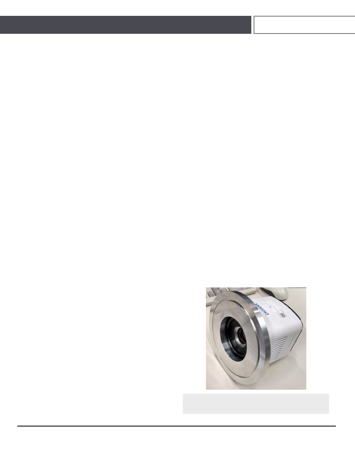
front side of the camera. The modified camera is depicted in Fig. 1.
The used CCD camera is the GE 2048 2048 from Greateyes
GmbH.28 Its back-illuminated chip is 27.6 ×27.6mm2large and has
a pixel size of 13.5 ×13.5 μm2. The quantum efficiency ranges from
0.65 at 284eV (C K-edge) to 0.8 at 1303eV (Mg K-edge). The gain
is 1 count/e−, and the full well capacity is 100 ke−. At −20○C and
1MHz readout speed (0.25fps), the camera features a readout noise
of 8.5 e−and a dark noise of 1 e−/pixel/s.
B. Experimental setup
In this work, a laser-produced plasma (LPP) was used as the
source of soft x-ray radiation. The source is explained in detail in
Ref. 29. Briefly, the LPP consists of a Yb:YAG thin disk laser with a
repetition rate of 100Hz and a solid state metal target. A laser pulse
with a wavelength of 1030nm and a pulse energy of up to 200 mJ is
focused onto a rotating cylinder with a changeable target material
(typically Cu or W), which forms a hot dense plasma with a
size in the range of 50 μm (FWHM). The pulse duration can be
continuously varied between 0.5ns and 30ns. The plasma
emits polychromatic and isotropic radiation in the range of
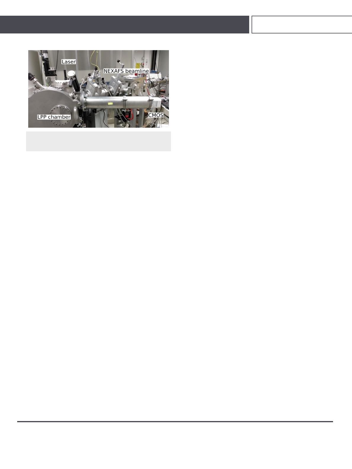
100 eV–1600eV. A photograph of the setup is shown in Fig. 2.
The XAFS measurements were performed using a twin-arm
reflection zone plate spectrometer previously described in Ref. 22.
The sample is measured in transmission mode, and the incident
x-ray spectrum is collected simultaneously using a pair of off-
axis reflection zone plate optics. A 200nm thick Al filter blocks
FIG. 1. Photograph of the Tucsen Dhyana 95 sCMOS camera that was modified
for the use in the soft x-ray range. The entrance window was removed and an
ISO–K 160 vacuum flange was attached.
Rev. Sci. Instrum. 92, 023102 (2021); doi: 10.1063/5.0032628 92, 023102-2
Published under license by AIP Publishing
Review of
Scientific Instruments ARTICLE scitation.org/journal/rsi
FIG. 2. Photograph of the setup including the Yb:YAG thin disk laser, the LPP
chamber, and the two beamlines used for the characterization measurements
(front) and the XAFS measurements (back).
residuallaser light, thevisiblepartof theemissionspectrumand sec-
ond order diffraction. The dispersed light is collected using either
the sCMOS or the CCD detector. The CCD is cooled down to
−30○C, and the sCMOS chip is operated at T =−15○C for all pre-
sentedmeasurements. Theabsorption ofthesampleisthen obtained
by the natural logarithm of the incident x-ray spectra divided by
the sample spectra. Through the use of multiple pairs of zone
plates, all existing absorption edges between 284eV (C K-edge) and
1303eV (Mg K-edge) can be measured with a resolving power
of E/ΔE≥900. The resolving power of the spectrometer at the
Ni L-edges amounts to roughly 1000. The whole spectrometer is
under high vacuum conditions (10−6mbar).
For the characterization of the sCMOS, the camera was
mounted to the chamber in 110cm distance to the target. The gain
was determined using monochromatized radiation and the single
photon counting procedure. To attenuate the produced radiation
to a photon flux suitable for single photon counting, four layers of
aluminum foil of a total thickness of 52 μm were inserted between
the target and the detector. The copper plasma emission spectrum
convoluted with the transmission of the Al filters results in a mean
photon energy of Emean =(1440 ±30) eV.
The gain was measured via single photon counting. Frames
with 10ms exposure time were recorded continuously with the LPP
at a repetition rate of 100Hz so that one shot was recorded on each
frame on average. 200 frames and 300 dark frames were recorded
for each measurement. The median of all dark frames was sub-
tracted, and for each pixel, the standard deviation was calculated.
Thestandarddeviation values were then usedtodeterminethenoise
thresholds for each pixel. The algorithm used to detect photon
events on the frames and to assign an intensity value in analog-to-
digitalunits (ADU)wastheclusteringalgorithm describedinRef.30
with threshold values of σ1=6 and σ2=3. To determine an accurate
result for the camera gain, only one-pixel events were evaluated.
The sCMOS dark current was measured by recording dark
frames for exposure times between 1s and 10s, with 20 frames for
each measuring point. Since the detector shows some regions with
unusually high dark current at the edges of the chip, only a section
of the total frames was evaluated to avoid distortion of the results.
This value can be seen as a lower limit for the dark current, while
an upper limit is provided by evaluating the whole frame. The mean
values of the selected region were plotted over exposure time and
the dark current was obtained from the slope of the linear fit. The
actual fit error was slightly raised to one significant digit to account
for possible small temperature fluctuations.
The readout noise was measured by taking five times 500 dark
frames with 20 μs exposure time each, so that the dark noise is neg-
ligible. The standard deviation from 500 dark frames was calculated
for each pixel and averaged for the whole frame. For the five mea-
surement series, the mean value was calculated. Since the standard
errors for both HG and HDR modes are smaller than the difference
of both mean values, the errors were estimated on the basis of this
difference.
The noise contribution originating from differing offset values
ofthepixels,thedarksignal non-uniformity (DSNU), was measured
with the same set of dark frames. For each set of 500 dark frames,
the mean value for each pixel was calculated and from those val-
ues the DSNU was determined by calculating the standard deviation
of the mean pixel values and subtracting the temporal noise. This
step was repeated for each set of 500 frames, and the mean value
and thestandard error were calculated from the five sets. Prior mea-
surements have shown differing values outside of the calculated
error range, so a greater error was estimated based on the difference
of the results.
For measuring the gain dependent part of the overall noise
originating from the different gain characteristics of the readout
amplifiers (pixel response non-uniformity, PRNU), flat field frames
are necessary, where the sCMOS chip is homogeneously illuminated
to investigate the different pixel responses. As a simple alternative
to an Ulbricht sphere, a gooseneck lamp combined with a pinhole
as the source of homogeneous illumination in the visible range was
used for the flat field frames. Frames were recorded for exposure
times between 100ms (10% saturation) and 250ms (50% satura-
tion) in 50ms steps in the HDR mode. For every measuring point,
500flatfieldframesinadditionto250darkframesweretaken,which
were averaged and subtracted from the flat field frames to mitigate
the DSNU. The relative PRNU was estimated by dividing the frame
standard deviation by the mean frame value. A homogeneous part
of the frame was chosen to obtain a lower estimate for the PRNU,
while the whole frame was evaluated for an upper estimate (assum-
ing some degree of non-uniformity in the illumination). The mean
values of the four measuring points were calculated. For the opera-
tionofthesCMOScamera,themanufacturer’ssoftwareMosaic1.6.1
was used.
C. Samples
For the XAFS measurements, a Ni sample has been fabri-
cated by resistive thermal evaporation at the nanoscale lab of the
Max–Born-Institute (MBI). The sample consists of a thin Ni layer
with a thickness of 100nm on top of a 150nm thick Si3N4window
with a size of 3 ×3 mm2.
As another example, a sandwich sample consisting of three lay-
ers (TiO2on Au-nanoparticles on NiO) was measured at the Ni
L-edges. The exact layer thicknesses are unknown but it is expected
that they amount to some tens of nanometers. The sample was
prepared on a 150nm thick, 1 ×2mm2Si3N4window. The sam-
ple system finds application as a plasmonic solar cell prototype
device.31,32
Rev. Sci. Instrum. 92, 023102 (2021); doi: 10.1063/5.0032628 92, 023102-3
Published under license by AIP Publishing
Loading more pages...