
Received: 12 July 2022 Revised: 25 August 2022 Accepted: 12 September 2022
DOI: 10.1002/nano.202200154
RESEARCH ARTICLE
Formation of diffraction gratings in optically patternable
hydrogel films
Tina Sabel-Grau Zhenfang Zhang Rahima Rahman Marga C. Lensen
Nanopatterned Biomaterials (Secr. C 1),
Department of Chemistry, Technische
Universität Berlin, Berlin, Germany
Correspondence
Tina Sabel-Grau and Marga C. Lensen,
Nanopatterned Biomaterials (Secr. C 1),
Department of Chemistry, Technische
Universität Berlin, Straße des 17. Juni 115,
10623 Berlin, Germany.
Email: [email protected];
Marga@lensenlab.de
Abstract
Local light-induced material response forms the basis for volume holographic
patterning of light-responsive materials. This enables applications as multifunc-
tional biomedical materials capable of integrating optical functions, such as for
advanced intraocular lens (IOL) implants. Therefore, hydrogel films based on 8-
arm PEG and azobenzene-functionalized acrylates are prepared. A local change
in optical properties is induced by UV exposure: a decrease in refractive index is
detected by ellipsometric examination. One-dimensional diffractive surface grat-
ings, generated using a photomask, show optical functionality and are visualized
in the atomic force microscope. The results show that a local change in opti-
cal properties can be induced in functionalized hydrogel films by photochemical
crosslinking, making them suitable for volume holographic patterning.
KEYWORDS
diffraction gratings, hydrogels, light-responsive materials, multifunctional biomedical bioma-
terials, photo curing, photopolymers, volume holography
1 INTRODUCTION
Multifunctional biomedical materials capable of integrat-
ing optical functions are highly desirable for many applica-
tions, such as advanced intraocular lens (IOL) implants.[1]
In the case of intraocular lenses, diffractive structures
are already being used at the current stage of development
to improve the optical functionality of the implant.[2]So
far, only surface structuring has been used. Optically struc-
turing the volume of the IOL – whether in addition or
instead of surface patterning – is an as yet unexploited,
but nevertheless great advancement. However, new light-
sensitive biomaterials are needed to exploit this potential.
Advanced IOL implants are only one example why
new materials for volume holography are constantly being
developed and researched with relentless intensity.[3,4]
This is an open access article under the terms of the Creative Commons Attribution License, which permits use, distribution and reproduction in any medium, provided the
original work is properly cited.
© 2022 The Authors. Nano Select published by Wiley-VCH GmbH.
Most photosensitive materials are designed either for
volume or for surface patterning. However, some systems
have been described for hybrid volume/surface gratings,
where a periodic modulation of the surface is observed in
addition to a volume phase grating.[5–7]Such dual grating
structures are of exceptional interest for many applications
and offer special opportunities for a deeper understanding
of the underlying grating formation mechanisms.[8]
As a volume holographic material, photosensitive poly-
mers represent a particularly interesting group among
stimuli-responsive polymeric materials, characterized by
their ability to be used in a non-invasive and eas-
ily controllable manner.[9]Light as a stimulus enables
optical patterning by applying volume holography as a
single-step method to fabricate diffractive 3D micro- and
nanostructures.[10]
Nano Select 2022;3:1583–1587. wileyonlinelibrary.com/journal/nano 1583
26884011, 2022, 12, Downloaded from https://onlinelibrary.wiley.com/doi/10.1002/nano.202200154 by Cochrane Germany, Wiley Online Library on [20/02/2023]. See the Terms and Conditions (https://onlinelibrary.wiley.com/terms-and-conditions) on Wiley Online Library for rules of use; OA articles are governed by the applicable Creative Commons License

1584 SABEL-GRAU et al.
The formation of holographic gratings in polymers
is determined by the interplay of polymerization and
diffusion – where the chemical gradient is always the driv-
ing force: If a component is consumed (e.g., by bonding
and crosslinking) or changed (e.g., by isomerization), a
concentration gradient is created. This results in diffusion
of the consumed or changed components and is accompa-
nied by a material transport, which creates the permanent
grating.[11]
While light-induced polymerization-diffusion processes
enable the structuring of any optically transparent, pho-
tosensitive material that exhibits sufficient mobility in
the sense of diffusion, permanent structures can only be
achieved if the diffusing species are incorporated into a
network.
Employing photosensitive hydrogels combines the
advantages of hydrogels and light; the hydrated structure
of the hydrogel makes them favorable as a bio-friendly
material, and the use of light as a patterning tool allows
dynamic control of their properties. Combination of these
advantages makes these systems attractive for biomedical
applications.[12]
Hydrogels, for example, based on poly(ethylene glycol)
(PEG), are generally highly interesting for application as
scaffolds for tissue engineering. They are cytocompatible
and intrinsically resistant to protein and cell adhesion, and
they can be controlled and functionalized on the molecular
level.[13]Numerous applications open up based on explicit
control over molecular structure and mechanical proper-
ties, such as elasticity and degree of crosslinking, as well
as surface morphologies.[14]
As biomaterials, hydrogels offer versatile possibilities
for (photo)chemical crosslinking and are suitable for the
generation of topographic surface structures as well as
for three-dimensional, macroporous scaffolds. In addition,
hydrogels can also be structured photolithographically
relying on diffusion processes.[15]The latter phenomenon
forms the basis for volume holography.
Photochromic hydrogels exhibit excellent reversible
conversion behavior, which can also be used for repeatable
writing of optical information.[16]Photomechanical hydro-
gels based on typical molecular photoswitches – such as
azobenzene – function as smart materials that respond to
light.[17]
The azobenzene moieties switch from trans to cis
conformation upon exposure to UV light and reverse
their conformation upon exposure to the visible light.
Also, these gels proved to be thermally responsive as
well. Thermal cis→trans isomerization occurs sponta-
neously due to the thermodynamic stability of the
trans isomer.[18]Thereby, in case of azobenzene-based
light-driven real-time information-transmitting systems,
the information is expected to be transmitted at the
molecular scale with response times ultimately within
the nanosecond or picosecond range.[19]In this pro-
cess, azo-dye molecules can also be used as diffus-
ing dopants to create holographic gratings.[20]Subwave-
length surface relief and refractive index gratings can
be recorded in azobenzene-containing liquid-crystalline
polymer films.[21]Multiresponsive supramolecular liquid-
crystalline polymer organogels are suitable for the inscrip-
tion of holographic gratings with switching behavior.[22]
Recent work with functionalized hydrogels based on
8-arm PEG has shown that the mechanical and physic-
ochemical properties can be controlled by the degree
of crosslinking, with uncrosslinked functional groups
remaining after gel formation.[23]The cytocompatibil-
ity of the material was positively evaluated. Such a
(bio)chemically functionalized photosensitive gel is partic-
ularly suitable for applications with specific biointeraction
requirements.
2 RESULTS AND DISCUSSION:
LIGHT-INDUCED STRUCTURING OF THE
HYDROGEL LAYERS
2.1 Local change in optical properties
The refractive index of the film was determined by ellipso-
metric measurement. Results are shown in Table 1.
A comparison of exposed and unexposed areas shows
a decrease in refractive index due to UV exposure of the
order of 10−3in case of functionalized hydrogel. In addi-
tion, a dependence of the contrast on the azo content was
found: An azo content of 0.3% results in a contrast of 0.005.
A higher azo content of 0.6% shows a higher contrast of
0.007.
The change in optical properties found can be attributed
to transformation processes in the molecular structure.
UV exposure causes the material to crosslink, whereby
double bonds are converted to single bonds. Single bonds
have a lower polarizability compared to double bonds.
The transformation should therefore be accompanied by
a reduction of the refractive index. On the other hand, the
crosslinking arguably causes an increase in the density of
the material, which is linked to an increase in the refractive
index. Experimentally, a decrease in refractive index was
observed in the course of UV exposure-induced crosslink-
ing. Thus, the increase in polarizability seems to outweigh
the increase in density of the material.
2.2 Generation of one-dimensional
diffractive surface gratings
The functionalized hydrogel layers were UV-crosslinked
under a photomask (1D modulated; lattice constant
26884011, 2022, 12, Downloaded from https://onlinelibrary.wiley.com/doi/10.1002/nano.202200154 by Cochrane Germany, Wiley Online Library on [20/02/2023]. See the Terms and Conditions (https://onlinelibrary.wiley.com/terms-and-conditions) on Wiley Online Library for rules of use; OA articles are governed by the applicable Creative Commons License
SABEL-GRAU et al. 1585
TABLE 1 Elypsometrically determined local refractive index n of the hydrogel layers
Refractive index Sample A (without Azo) Sample B (0.3% Azo) Sample C (0.6% Azo)
Unexposed 1.417 ±0.006 1.423 ±0.002 1.429 ±0.002
UV exposed 1.423 ±0.008 1.418 ±0.002 1.422 ±0.002
Contrast −0.005 −0.007
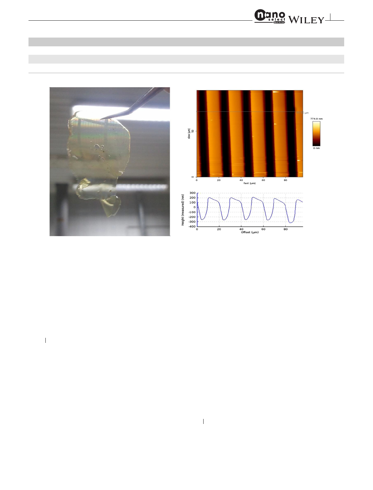
FIGURE 1 Diffractive structures in hydrogel: hydrogel film with optical functionality (left) and corresponding surface topography (right)
20 µm). In this process, diffractive surface gratings were
formed. Figure 1shows a layer with optical functionality,
as well as the corresponding surface characteristics (topog-
raphy profile from atomic force microscopy studies) of the
patterned hydrogel. In general, detailed analysis of SRG
topography provides a good opportunity to gain insight
into the grating formation processes.[8]
2.3 Grating formation in functionalized
hydrogel
Among the various effects that can contribute to grat-
ing formation via a local change in optical properties are:
basically, the exposure-induced change in refractive index
due to crosslinking of acrylate groups and the change in
density due to crosslinking. In the case of azo compo-
nents, the cis/trans isomerization causes local changes
in molecular structures or configurations, in connection
with changes in polarizability with direct effects on the
refractive index.[24]In addition, cis→trans isomerization
can also contribute to a change in density.[25]Then the
important aspect of photo-triggered mass migration in the
cause of component diffusion is to be considered: both with
respect to diffusion of hydrogel components PEG[15]and
with respect to diffusion of the azo-dye.[20]
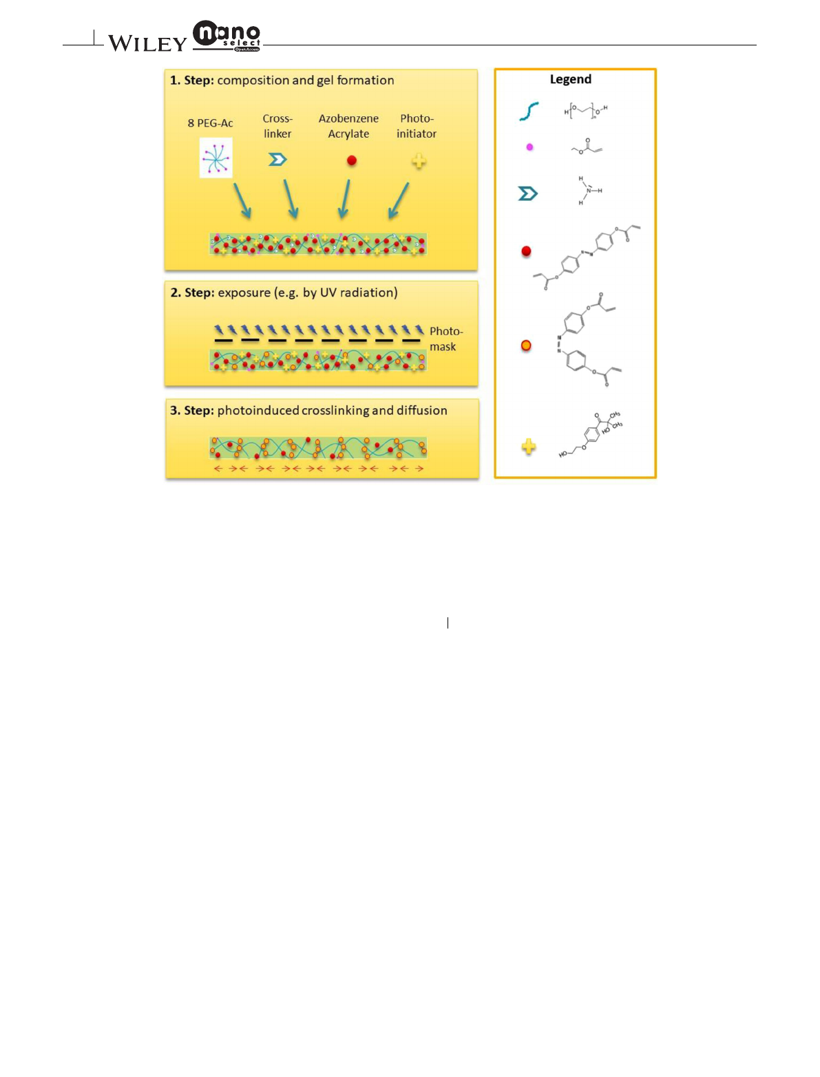
The anticipated grating formation in functionalized
hydrogel is schematically shown in Figure 2.
In a first step, the mixture forms a gel by reaction of
8PEG and crosslinker (ammonia), leaving uncrosslinked
functional groups (acrylate groups). In the second step,
the structured exposure is performed by a photomask (or,
if volume holographic structuring is used, by an interfer-
ence pattern). The photoinitiator is consumed, with pho-
toinduced attachment of the dopant (here, azobenzene-
functionalized acrylates). In addition, trans→cis isomer-
ization occurs under UV irradiation. The third step, dif-
fusion of uncrosslinked dopant (and possibly – depending
on the exposure dose – also crosslinking and isomerization
of the diffused components), forms the permanent optical
grating.
3 CONCLUSION
It was shown that a local change in optical properties
can be induced in special functionalized hydrogel films
by photochemical crosslinking. A compound containing
26884011, 2022, 12, Downloaded from https://onlinelibrary.wiley.com/doi/10.1002/nano.202200154 by Cochrane Germany, Wiley Online Library on [20/02/2023]. See the Terms and Conditions (https://onlinelibrary.wiley.com/terms-and-conditions) on Wiley Online Library for rules of use; OA articles are governed by the applicable Creative Commons License
1586 SABEL-GRAU et al.
FIGURE 2 Anticipated grating formation in functionalized hydrogel
8-PEG and azo was used, with the advantage of multiple
crosslinking points that allow the azo units to form the gel.
We found a decrease in refractive index over the course
of UV irradiation-induced crosslinking, which can be
attributed to the increase in polarizability, and which
seems to outweigh the increase in density of the material.
As we know, an inducible refractive index contrast of
the order of 10−3allows a diffraction efficiency close to
100% for 200 µm thick layers.[11]It should be noted that
the experiments performed here to determine the refrac-
tive index contrast do not even involve diffusion processes.
Thus, the contrast is not due to material transport, but only
due to changes in molecular structure. Since azo compo-
nents are involved here, trans→cis isomerization is also
a possibility for the contrast-effective change in molecu-
lar structure.[24]However, by integrating polymerization-
diffusion processes in the form of holographic patterning,
a much higher contrast can therefore be expected.
One-dimensional diffractive surface gratings were cre-
ated using a photomask. Such structured hydrogel layers
show optical functionality, and the related surface topog-
raphy was visualized in the atomic force microscope.
The observed optical functionality may be caused by the
measured surface grating, or additionally by a possibly
generated correlated volume phase grating. Which con-
tribution each of the two have, must be elucidated by
further investigations, preferably by holographic exposure
and exact analysis of the diffraction properties.
4EXPERIMENTAL SECTION –
MATERIALS AND METHODS
A composition based on an 8-arm star-shaped PEG with
acrylate groups (8PEG) was chosen for the preparation of
the hydrogel film, and an azo compound (azobenzene-
functionalized acrylates) was added.[18]
This mixture was placed between two glass slides with
the addition of photoinitiator (2%), ammonia (30%), and
water (100%). Ammonia acts as a crosslinking agent and
induces the gel formation.[23]After gel formation with
NH3, the gels were exposed to UV light (366 nm) for 5–7
minutes. The cured gels were peeled off and the samples
were placed on clean glass slide. The solvent was evap-
orated and further dried until constant weight. The film
thickness was 100 µm. The refractive index of the film was
then determined ellipsometrically.
ACKNOWLEDGMENTS
This research was funded by Deutsche Forschungsge-
meinschaft (DFG, German Research Foundation), grant
number SA 2990/1-1.
26884011, 2022, 12, Downloaded from https://onlinelibrary.wiley.com/doi/10.1002/nano.202200154 by Cochrane Germany, Wiley Online Library on [20/02/2023]. See the Terms and Conditions (https://onlinelibrary.wiley.com/terms-and-conditions) on Wiley Online Library for rules of use; OA articles are governed by the applicable Creative Commons License

SABEL-GRAU et al. 1587
Open access funding enabled and organized by Projekt
DEAL.
CONFLICT OF INTEREST
The authors declare no conflict of interest.
DATA AVAILABILITY STATEMENT
The data that support the findings of this study are
available from the corresponding author upon reasonable
request.
REFERENCES
1. T. Sabel-Grau, A. Tyushina, C. Babalik, M C. Lensen, Gels.2022,
8(164), 8.
2. X.Xu,M.M.Zhu,H.D.Zou,J. Refract. Surg.2014,30(9), 634.
3. J. Guo, M. R. Gleeson, J. T. Sheridan, Phys. Res. Int.2012,2012,
16.
4. V. A. Barachevsky, Opt. Spectrosc.2018,124,373.
5. N. D. Vorzobova, P. P. Sokolov, V. O. Veselov, I J. Schelkanova,
Appl. Opt. 2018,57(12), 3323. Available from: http://ao.osa.org/
abstract.cfm?URI=ao-57-12-3323
6. I. Naydenova, E. Mihaylova, S. Martin, V. Toal, Opt. Express.
2005,13(13), 4878.
7. D. E. Lucchetta, P. Spegni, A. Di Donato, R. Castagna, Opt. Mater.
(Amst),2015,42, 366.
8. T. Sabel, V. de G. Lucas, M. C. Lensen, Mater. Res. Lett.2019,
7(10), 405.
9. D. Roy, J. N. Cambre, B. S. Sumerlin, Prog. Polym. Sci.
2010,35(1), 278. Available from: http://www.sciencedirect.com/
science/article/pii/S0079670009001063
10. W. S. Colburn, K. A. Haines, Appl. Opt.1971,10(7), 1636. Avail-
able from: http://ao.osa.org/abstract.cfm?URI=ao-10-7-1636
11. T. Sabel, M C. Lensen, Holographic Materials and Optical Sys-
tems (Eds: I. Naydenova, T. Babeva, D. Nazarova), InTech,
Rijeka, Croatia 2017.
12. I. Tomatsu, K. Peng, A. Kros, Adv. Drug. Deliv. Rev.2011,63(14),
1257. Available from: https://www.sciencedirect.com/science/
article/pii/S0169409%D7;11001542
13. K.T.Nguyen,J.L.West,Biomaterials 2002,23(22), 4307.
14. M. C. Lensen, V. Schulte, J. Salber, M. Dietz, F. Menges, M.
Möller, Pure. Appl. Chem.2008,80(11), 2479.
15. B. M. S. Hahn, J. S. Miller, J. L. West, Adv. Mater.2006,18,2679.
16.S.Long,Z.Ye,Y.Jin,J.Huang,Y.Huang,Y.Liao,etal.
Macromol. Rapid. Commun.2021,42(7), 2000701. Available
from: https://onlinelibrary.wiley.com/doi/abs/10.1002/marc.
202000701
17. P.F.Luo,S.L.Xiang,C.Li,M.Q.Zhu,J. Polym. Sci.2021,59(20),
2246. Available from: https://onlinelibrary.wiley.com/doi/abs/
10.1002/pol.20210567
18. Rahman R. Photo - and Thermo - Responsive Poly (Ethylene Gly-
col) - based Biomaterials: Synthesis, Characterization, Pattern-
ing and Application in Biological Studies [Internet]. Technische
Universität Berlin; 2018. Available from: https://depositonce.tu-
berlin.de/bitstream/11303/7762/4/rahman_rahima.pdf
19. J. García-amorós, D. Velasco, Beilstein. J. Org. Chem.2012,8,
1003.
20. C.T.Kuo,S.Y.Huang,Appl. Phys. B.2007,86(4), 699. Available
from: https://doi.org/10.1007/s00340-006-2562-x
21. H. Yu, A. Shishido, T. Ikeda, Appl. Phys. Lett.2008,92(10), 103117.
Available from: https://doi.org/10.1063/1.2894073
22. Y. Ni, X. Li, J. Hu, S. Huang, H. Yu, Chem. Mater.2019,31(9),
3388. Available from: https://doi.org/10.1021/acs.chemmater.
9b00551
23.Z.Zhang,A.Loebus,G.deVicente,F.Ren,M.Arafeh,Z.
Ouyang, et al. Chem. Mater.2014,26(12), 3624. Available from:
https://doi.org/10.1021/cm500203j
24. Y.J.Liu,H.T.Dai,E.S.P.Leong,J.H.Teng,X.W.Sun,Opt.
Mater. Express.2012,2(1), 1667.
25. E. Merino, M. Ribagorda, Beilstein. J. Org. Chem.2012,8,1071.
How to cite this article: T. Sabel-Grau, Z. Zhang,
R. Rahman, M. Lensen, Nano Select.2022,3, 1583.
https://doi.org/10.1002/nano.202200154
26884011, 2022, 12, Downloaded from https://onlinelibrary.wiley.com/doi/10.1002/nano.202200154 by Cochrane Germany, Wiley Online Library on [20/02/2023]. See the Terms and Conditions (https://onlinelibrary.wiley.com/terms-and-conditions) on Wiley Online Library for rules of use; OA articles are governed by the applicable Creative Commons License
Loading more pages...