New J. Phys. 24 (2022) 113043 https://doi.org/10.1088/1367-2630/aca176
OPEN ACCESS
RECEIVED
6 May 2022
REVISED
31 October 2022
ACCEPTED FOR PUBLICATION
9 November 2022
PUBLISHED
30 November 2022
Original content from
this work may be used
under the terms of the
Creative Commons
Attribution 4.0 licence.
Any further distribution
of this work must
maintain attribution to
the author(s) and the
title of the work, journal
citation and DOI.
PAPER
Diffraction imaging of light induced dynamics in xenon-doped
helium nanodroplets
BLangbehn
1,∗,YOvcharenko
1,2,AClark
3,MCoreno
4,RCucini
5,
A Demidovich6, M Drabbels3, P Finetti6,MDiFraia
4,6, L Giannessi6, C Grazioli5,
DIablonskyi
7,ACLaForge
8, T Nishiyama9, V Oliver ´
Alvarez de Lara3,CPeltz
10,
PPiseri
11,OPlekan
6,KSander
10,KUeda
7, T Fennel10,12 ,KCPrince
6,13 ,
FStienkemeier
8, C Callegari4,6,TM¨
oller1and D Rupp12,14
1Technische Universit¨
at Berlin, Institut f¨
ur Optik und Atomare Physik, 10623 Berlin, Germany
2European XFEL GmbH, 22607 Hamburg, Germany
3Laboratory of Molecular Nanodynamics, Ecole Polytechnique F´
ed´
erale de Lausanne (EPFL), 1015 Lausanne, Switzerland
4ISM-CNR, Istituto di Struttura della Materia, LD2 Unit, 34149 Trieste, Italy
5IOM-CNR, Istituto Officina dei Materiali, 34149 Trieste, Italy
6Elettra–Sincrotrone Trieste, S.C.p.A., 34149 Trieste, Italy
7Institute of Multidisciplinary Research for Advanced Materials, Tohoku University, Sendai 980-8577, Japan
8Institute of Physics, University of Freiburg, 79104 Freiburg, Germany
9Division of Physics and Astronomy, Graduate School of Science, Kyoto University, Kyoto 606-8502, Japan
10 Institut f¨
ur Physik, Universit¨
at Rostock, 18059 Rostock, Germany
11 CIMAINA and Dipartimento di Fisica, Universit`
a degli Studi di Milano, 20133 Milano, Italy
12 Max-Born-Institut f¨
ur Nichtlineare Optik und Kurzzeitspektroskopie, 12489 Berlin, Germany
13 Department of Chemistry and Biotechnology, Swinburne University of Technology, Victoria 3122, Australia
14 Laboratorium f¨
ur Festk¨
orperphysik, ETH Z¨
urich, 8093 Z¨
urich, Switzerland
∗Author to whom any correspondence should be addressed.
E-mail: bruno.langbehn@physik.tu-berlin.de,thomas.moeller@physik.tu-berlin.de and
Keywords: coherent diffraction imaging, helium nanodroplets, superfluid, dynamics
Abstract
We explore the light induced dynamics in superfluid helium nanodroplets with wide-angle
scattering in a pump–probe measurement scheme. The droplets are doped with xenon atoms to
facilitate the ignition of a nanoplasma through irradiation with near-infrared laser pulses. After a
variable time delay of up to 800 ps, we image the subsequent dynamics using intense extreme
ultraviolet pulses from the FERMI free-electron laser. The recorded scattering images exhibit
complex intensity fluctuations that are categorized based on their characteristic features.
Systematic simulations of wide-angle diffraction patterns are performed, which can qualitatively
explain the observed features by employing model shapes with both randomly distributed as well
as structured, symmetric distortions. This points to a connection between the dynamics and the
positions of the dopants in the droplets. In particular, the structured fluctuations might be
governed by an underlying array of quantized vortices in the superfluid droplet as has been
observed in previous small-angle diffraction experiments. Our results provide a basis for further
investigations of dopant–droplet interactions and associated heating mechanisms.
1. Introduction
The interaction of intense laser pulses with matter plays an important role in many fields, from modern
high-tech applications such as laser machining [1]andmicrosurgery[2], extreme ultraviolet (XUV)
lithography [3], or propulsion of small satellites [4,5] to fundamental and applied science. For example, in
structure analysis of biological specimen using x-ray diffraction, radiation damage poses a substantial
problem, thus fueling a considerable research interest, in the processes accompanying and following the
irradiation with intense x-ray bursts [6].
© 2022 The Author(s). Published by IOP Publishing Ltd on behalf of the Institute of Physics and Deutsche Physikalische Gesellschaft

New J. Phys. 24 (2022) 113043 BLangbehnet al
In particular, atomic clusters can serve as isolated model systems of simple geometrical structure.
Time-resolved studies, where an initial pump light pulse excites the system and a subsequent probe light
pulse measures the state of the system after a defined temporal delay, enable observation of the light
induced dynamics. In the extreme case, the free cluster will eventually fully disintegrate, as the energy
deposited by the pump pulse cannot dissipate otherwise. A detailed investigation of the fragments and the
dynamics can give an insight into the processes and their inherent time scale, which, in comparison with
theoretical models of the interaction, enable development of a fundamental understanding [7–12]. A basic
picture divides the interaction of an intense light pulse with an atomic cluster into three steps [13,14]:
(i) the ionization of cluster atoms and emission of electrons until the increasing positive charge prevents
further electrons from leaving the cluster, (ii) the formation and evolution of a nanoplasma of ions and
quasi-free electrons on a short timescale, often only a few femtoseconds, and (iii) an expansion leading to
complete destruction of the cluster, accompanied by relaxation and recombination processes.
Helium clusters, often referred to as helium nanodroplets, are model systems particularly suited to such
studies due to their very simple electronic structure and smooth behavior: they remain liquid down to
absolute zero forming mostly spherical shapes [15,16], and they are transparent from the far-infrared to the
vacuum ultraviolet because of the large ionization potential of helium (24.6 eV). Using a near infrared
(NIR) laser pulse, helium nanodroplets can be ionized only when the power density is sufficiently high that
multiphoton ionization or field driven processes like tunneling ionization become significant
(∼1015 Wcm
−2)[17,18]. Another option is to dope the droplets using atoms with a low ionization
potential: in this case, ionization becomes possible already at lower laser intensity [19,20], starting an
avalanche-like process at the dopant positions [21,22]. Depending on the interaction of the impurity with
the helium solvent, the dopants remain at the surface of the droplet or get immersed, as is the case for, e.g.,
xenon atoms [23]. When xenon-doped droplets are irradiated by an NIR pulse, theory predicts a very
efficient complete ionization even with only a few xenon atoms embedded [19]. For the simple model of a
compact dopant cluster in the center of the helium droplet, an anisotropic growth of the nanoplasma along
the laser polarization axis has been predicted upon ignition [19]. On the other hand, dopant cluster
formation has been observed at multiple centers in larger droplets [24]. Further, the superfluid nature of the
helium nanodroplets leads to the occurrence of quantized vortices in large droplets [15], and the dopants
agglomerate along the vortex lines [25,26]. Nanoplasma ignition can therefore be expected in large droplets
at multiple sites, randomly distributed or structured by the vortex positions, which might lead to complex
dynamics. In addition to nanoplasma dynamics governed by Coulombic forces between charged
constituents, geometric changes beyond homogeneous Coulombic explosion or hydrodynamic expansion
might have to be considered in helium nanodroplets. For example, the formation of voids around free
electrons, so-called electron bubbles, has been described in liquid helium [27]. Further, the formation of gas
bubbles has been observed on a macroscopic scale around copper nanoparticles in bulk liquid helium [28]
and has also been reported on a microscopic scale around silver clusters in helium nanodroplets [29], in
both cases induced by resonant heating of the metal particles with an optical laser. Although superfluid
helium is known for its very large heat conductivity, the flow of thermal energy from a solid particle to
liquid helium is hindered by a discrepancy in velocity of sound, an effect called Kapitza resistance [29,30],
that leads to a sudden evaporation of the surrounding helium, eventually forming a gas bubble. A third
process that could be taken into account is the ejection of dopant ions from a helium nanodroplet, leaving
the droplet with several helium atoms attached to them [31,32]. These considerations show that the study
of light induced dynamics in doped helium droplets may have particularly interesting aspects. So far, most
theoretical work has been carried out for relatively small helium droplets (N⩽106atoms per droplet).
While imaging experiments inherently address larger droplets [15,16], their outcome has already stimulated
theoretical studies, e.g., on the droplet shapes [33,34]. It has been the close interplay between theory and
experiment that established a more profound knowledge of the underlying physics of rotating superfluid
droplets [35]. Therefore, it would be particularly interesting to experimentally visualize the droplet
dynamics after laser excitation, giving a basis for comparison with theoretical models. Eventually, if droplet
ignition at multiple sites or geometric changes in the droplets are observed, such experiments might lead to
an extension of the models or even to a new approach to gain further insight into the processes, e.g., by
employing molecular dynamics simulations.
In this work, we report on a study of the dynamics triggered by intense NIR laser pulses in large,
xenon-doped helium nanodroplets. In this context, the advent of short-wavelength free-electron lasers
(FELs) has opened up a new route to determine the structure of individual nanoparticles via coherent
diffraction imaging (CDI) [36], which is based on recording the light scattered off a particle. In general,
such a diffraction pattern reflects the scattering behavior of the particle, i.e., it depends on the distribution of
atoms and their response to the wavelength of the incident light. Therefore, a change in scattering strength,
e.g., because of a modification of the particle’s density or its electron distribution, leads to a change of the
2

New J. Phys. 24 (2022) 113043 BLangbehnet al
intensity distribution in the diffraction pattern, which is the basis for our method. The intense femtosecond
FEL light pulses enable the taking of a snapshot of the particle in time, thus allowing tracking of the
evolution of the particle in a pump–probe measurement scheme. This technique has been recently used to
study light induced dynamics in a variety of systems, e.g., structural changes in rare gas clusters [37–40],
surface melting of metal clusters [41], plasma dynamics in SiO2nanospheres [42], and anisotropic
evaporation of helium nanodroplets [18]. Here, we present a scenario where the energy is coupled into the
system via the dopants at localized positions. The agreement between outstanding features in our recorded
diffraction patterns and our qualitative model of randomly distributed and ordered voids in the droplets
gives a strong indication that the dynamics is not homogeneous but starts at several distinct sites in the
droplets.
2. Methods
The investigation is based on the analysis of wide-angle scattering images recorded at the FERMI FEL in a
similar configuration to that previously described [16]. The experimental setup is described in section 2.1.
In section 2.2, the procedure to simulate wide-angle diffraction patterns in order to retrieve information on
the droplet dynamics from the recorded scattering images is presented.
2.1. Pump–probe imaging setup
The experiment was carried out in a pump–probe measurement scheme at the low density matter
end-station [43] of the FERMI FEL. The helium nanodroplets are first irradiated with an NIR pulse
(wavelength λNIR =780 nm, duration 90 fs) to trigger dynamics (pump) and subsequently, after a variable
time delay of up to 800 ps, imaged using a 90 fs long XUV pulse (probe). The NIR laser is focused to a
100 μmspot(1/e2diameter) resulting in a power density of INIR =9×10¹³ Wcm
−2.TheFEListunedto
either non-resonant or resonant photon energies (Eph =19.4eVor21.5 eV, respectively) delivering power
densities exceeding IFEL =3×10¹⁴ Wcm
−2at spot sizes of 9 ×13 μm2(FWHM) and smaller. Please note
that the focus size of the NIR laser is chosen to be larger than that of the FEL to ensure that each droplet is
hit by the NIR laser before being imaged with the FEL. The XUV light scattered off a helium droplet is
collected up to a maximum scattering angle θmax =30◦by a detector consisting of a circular microchannel
plate (MCP) stacked onto a phosphor screen [7] located 65 mm downstream of the interaction region.
Pictures of the amplified scattering images on the phosphor screen are taken via a 45◦mirror using a
sCMOS Andor Neo 5.5 camera. The MCP, phosphor screen, and mirror have centered holes to let the FEL
and NIR beams pass through.
The helium droplets are produced by supersonic expansion into vacuum of helium through a 100 μm
trumpet-shaped nozzle with half opening angle of 20◦at a stagnation pressure p0=80 bar and temperature
T0=5.4 K, yielding a measured droplet velocity of 320 m s−1[44] and a mean droplet radius of
R=400 nm (corresponding to a mean droplet size of N=6×10⁹atoms per droplet) as determined
from scattering images of approximately spherical droplets [44]. The droplets get doped with xenon by
traversing a 35.3 mm long gas cell, successively capturing ∼1.6×10⁶xenon atoms per droplet (0.3 %)
[23,45]. The kinetic energy deposited in the droplet by each captured atom leads to the evaporation of
roughly 500 helium atoms [45,46]. Hence, the average droplet radius can be calculated to shrink to about
R=380 nm [47], which matches well the measured average radius of doped droplets. Overall, the
dopant atoms constitute only a small fraction of the total count of droplet atoms. Thus, for the wavelengths
used in this experiment we can assume the recorded diffraction patterns are dominated by light scattered off
the helium atoms.
In the analysis presented here, 26 390 images exhibiting meaningful scattering signal have been included.
The visibility of the scattering signal has been improved in post processing of the data by subtracting the
straylight background. In addition, the images have been corrected for the uneven detector sensitivity (that
is, e.g., due to the 8◦bias angle of the MCP [48]) and an angle-dependent intensity correction (∝cos−3θ)
has been applied that accounts for the flat shape of the detection locus [49]. The thus corrected patterns are
made available via the CXI data bank [50] under the identifier (ID) 208 [51].
2.2. Wide-angle scattering simulations
In order to retrieve information on the droplet density distribution, the diffraction patterns of individual
droplets are analyzed. In the case of small-angle scattering (θ5◦) the droplet’s electron density projection
can be reconstructed from the diffraction pattern using a phase retrieval algorithm which is commonly
utilized in CDI experiments and has already been successfully applied to doped helium nanodroplets
[25,26].Inthecaseofwide-angle scattering, however, multiple projection planes contribute to the
diffraction pattern rendering the reconstruction of a single projected density using algorithms based on
3

New J. Phys. 24 (2022) 113043 BLangbehnet al
inverse 2D Fourier transform impossible. For the same reason, wide-angle scattering images contain
valuable information on the three-dimensional droplet shape and orientation, that can be retrieved by
matching simulated diffraction patterns to the experimental data, as has been shown for faceted silver
clusters [52]. This forward-fitting technique has further been employed to investigate the shapes of spinning
helium nanodroplets [16,53,54] and is adapted here to address density fluctuations inside the droplets via
systematic simulations. The choice of employing systematic simulations instead of directly deriving the
shape information from the diffraction patterns was enforced by the firm constraints of the data set. These
are (i) the large number of strongly varying complex patterns, impossible to manually analyze in an
appropriate manner to generate reliable statistics of the abundances, (ii) the nonlinear detector response
together with the wide-angle detection which prevent object reconstruction by iterative phase retrieval, and
(iii) the large range of complicated features from inhomogeneous densities in the evolved droplets that
could not be captured by generalized model shapes, which are needed for direct forward fitting.
For calculating the wide-angle scattering patterns, a fast algorithm based on the multi slice Fourier
transform (MSFT) [55] approach was used. While its approximations neglect multiple scattering events,
e.g., backscattering of the propagating electromagnetic field, material properties can be approximated via
effective optical parameters even for photon energies close to the resonance [44]. These parameters have
been determined by matching scattering simulations to diffraction patterns of spherical helium
nanodroplets and comparing the radial intensity distribution to simulations based on Mie theory [56]fora
variety of droplet sizes and photon energies ranging from 19.0eVto23.7eV[44].
3. Results
In the following, the measured scattering images are described and discussed. First, the emergence of
dynamic features in the patterns is exemplified, i.e., of features that are not observed in static (XUV only)
data. In section 3.1, the temporal evolution of the patterns is investigated, while in section 3.2 different
classes of patterns are identified based on their salient features.
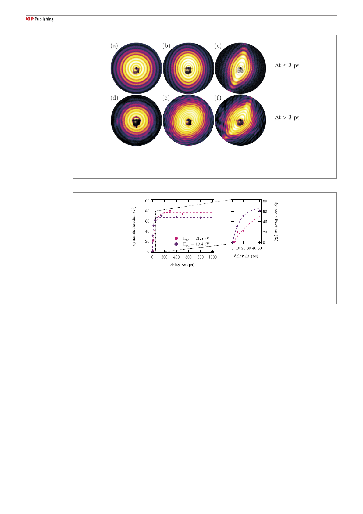
Figure 1shows a small selection of scattering images of xenon-doped helium nanodroplets at short time
delay Δtbetween pump and probe pulse. It should be noted that much more pronounced changes of the
scattering occur at longer delays which will be discussed later (cf figures 3and 4). In the early stages of the
dynamics, i.e., Δt⩽3 ps, see figures 1(a)–(c), the images exhibit no visible difference from those of pristine
droplets [16]: in contrast to data taken with hard x-rays [26] the dopants do not lead to a change of the
intensity distribution. Presumably, the nanometer-sized structures of dopant aggregates are not resolvable at
the wavelength of the incoming XUV pulse. However, as shown in figures 1(d)–(f), intensity fluctuations
along the diffraction rings become apparent for longer delays Δt>3 ps. We assume these fluctuations are
linked to the dynamics in the droplets and consequently refer to these data as images exhibiting dynamic
features. Details of the determination of the images showing dynamic features and an overview of the whole
data set are given in appendix A. Overall, for short time delays and only slight intensity fluctuations, the
droplet shapes can still be inferred from the known characteristic patterns [16] and classified as spherical
(figures 1(a) and (d)), ellipsoidal (figures 1(b) and (e)), and pill-shaped (figures 1(c) and (f)). Nonetheless,
the longer the delay the more complicated it is to identify droplet shapes, as the diffraction patterns become
increasingly distorted (cf figures 3and 4).
3.1. Temporal evolution of the patterns
In figure 2, the manually determined fraction of images exhibiting dynamic features is shown versus the
time delay Δtfor the non-resonant (Eph =19.4 eV) and resonant (Eph =21.5 eV) photon energy of the
XUV pulse. The reproducibility of the manually performed assignment of the dynamic fraction was tested
(see appendix A)andfoundtobebetterthan90%.Asaguidetotheeye,limitedgrowthfunctionsare
shown as dashed lines. Three main observations can be made: (i) there is a steep increase of the dynamic
fraction, that is faster for the non-resonant wavelength, (ii) the dynamic fraction saturates to less than 100%
at long delays (Δt>200 ps) indicating that not all images exhibit dynamic features, and (iii) the saturation
percentage is higher for the resonant wavelength.
We attribute the first point to the wavelength-dependent influence of the refractive index on the
scattering. In the resonant case (Eph =21.5 eV) the absorption is high, hence, mostly the front surface of
the droplet contributes to the scattering and density changes inside the droplet are not visible in the
diffraction pattern. When the dynamics initiated by the NIR pulse starts inside the droplet (which we expect
to be the case for xenon dopants) and propagates toward the surface, it can be observed earlier in the
diffraction patterns recorded at the non-resonant photon energy (Eph =19.4 eV). The second and third
observations, however, are most probably related to an experimental artifact: we assume that an imperfect
overlap of the pulses leads to a situation where the dynamics is obviously not initiated in every droplet, even
4
New J. Phys. 24 (2022) 113043 BLangbehnet al
Figure 1. Emergence of dynamic features in diffraction patterns of xenon-doped helium nanodroplets. (a)–(c) For a time delay
Δt⩽3 ps the scattering images resemble those of pristine droplets described before [16], i.e., the dopants do not lead per se to
visible changes in the patterns. (d)–(f) For longer delays, intensity fluctuations become apparent in the images that are linked to
the dynamics in the droplets and therefore referred to as dynamic features.
Figure 2. Temporal evolution of the fraction of scattering images exhibiting dynamic features. After a steep increase within
100 ps to 150 ps after NIR irradiation the dynamic fraction saturates at about 60% to 80%, i.e., the dynamics is not ignited in
every droplet. From the inset it can be seen that for short delays (Δt<3 ps) no dynamics are observed in the patterns. Further,
the rise of the dynamic fraction is slower for the resonant photon energy (Eph =21.5 eV), cf the data points at 20 ps. The dashed
lines are limited growth functions as a guide to the eye.
though a larger focal spot was chosen for the pump (NIR) than for the probe (XUV) pulse. Further, we
identify the third observation—a slightly lower asymptotic value for the non-resonant photon energy—as a
decreasing overlap between the two pulses over time, as even lower values were observed for subsequent
delay scans that were taken several hours apart (not shown).
The dynamic features in the patterns become, as expected, more pronounced with longer time delay. As
well, the intensity not only fluctuates along the diffraction rings, but the complexity of the intensity
distribution increases. It should be noted that many patterns exhibit strong directionality, which is
completely different from what was observed in earlier pump–probe experiments on pure rare gas clusters
[37,38], thus indicating multiple scattering centers at specific sites rather than surface smoothing or
homogeneous fragmentation of the particle. The development toward long time scales is exemplified in
figure 3, where the four brightest scattering images are shown for short (∼10 ps) and long (∼800 ps) delays
at non-resonant (figure 3(a)) and resonant (figure 3(b)) photon energy. It can be seen that the intensity
fluctuations at short delays are more pronounced for non-resonant scattering (Eph =19.4eV),wherethe
droplets are almost transparent and information on density fluctuations inside the droplets is encoded in
the diffraction pattern. For the resonant case (Eph =21.5 eV), less pronounced fluctuations presumably
reflect changes on the droplet surface that occur later in time. Note that this observation is in line with the
slower rise of the respective curve in figure 2.
The images shown in figure 3already exhibit complex features, especially at long delays. However, this
situation becomes even more complicated when not only the brightest images are examined but the whole
data set is analyzed. While the overall trend—the longer the delay, the more distorted the diffraction
pattern—remains the same, the degree of complexity in the images at a given delay varies considerably. In
particular, no definite evolution in time is discernible when individual patterns are compared. Two facts can
5
Loading more pages...